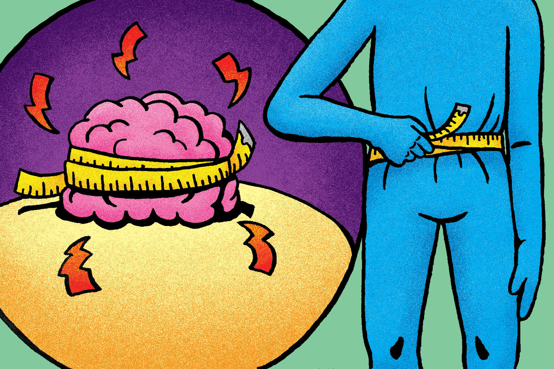
Expounding on Anorexia: Cognitive and Structural Outcomes
Author: Remya Brown || Scientific Reviewer: Charita Kunta || Lay Reviewer: Ashish Abraham || General Editor: Vishwanka Kuchibhatla || Artist: Ethan Carroll || Graduate Scientific Reviewer: Molly Tassoni
Publication Date: May 10, 2021

Anorexia nervosa (AN) is a psychiatric disorder with the highest mortality, mostly affecting young women in their adolescence [1]. While it generally takes five or six years to recover after an initial diagnosis, long-term interventions are difficult to maintain, and about 30% do not recover [1]. Because it can have damaging effects, people who have anorexia nervosa exhibit symptoms that may be apparent, such as extreme weight loss, thin appearance, and hair thinning [2]. These are some of the commonly recognized physical effects on one’s body. However, what is less known are some of the unseen effects on an individual. Recent studies have suggested that AN has detrimental effects on the structural and cognitive functions of the brain. Significant changes include a decrease in grey matter and mental flexibility. Scientists suggest that the repercussions of the eating disorder may or may not be reversible with recovery. As the goal of this article is to be informative, the following may be triggering content for readers who have or may be suffering from eating disorders.
AN is an eating disorder which mostly affects vulnerable individuals, primarily during their adolescence or young adulthood. The factors that increase the risk for AN can be genetic, dieting, starvation and major life changes that increase emotional wellbeing [2]. It is characterized by the intense fear of gaining weight or distorted perceptions of weight. Extreme efforts to control one’s weight can become unhealthy and maybe even life-threatening. In fact, AN is considered the third most common chronic disease among adolescents, as well as having the highest mortality rate out of many psychiatric disorders [3]. Individuals do manage to recover from the illness, however the long term repercussions are still in discussion. Cognitive flexibility, for example, is one of the few aspects of executive function that patients with AN appear to lack compared to their healthy counterparts, whereas most other abilities were comparable to one another for both groups [4]. In addition to the cognitive functions, AN has been linked to a reduced brain structure, especially grey matter, which may or may not be analogous to the clinical symptoms of the illness [5].

Impacts of AN on Cognitive Function
Inefficient cognition associated with AN has been identified in adults [4]. However, longitudinal research was necessary to determine how cognitive abilities change from adolescence to adulthood. In a study from Telléus and colleagues, the association between weight status and cognition following one year of recovery (or from the first time they were assessed) was analyzed. Originally, it was hypothesized that intelligence and cognitive function in individuals with AN would be significantly reduced compared to healthy individuals [4]. However, following one year of research, it was found that there was no significant difference in cognitive development between the two groups. Hence, in AN and recovery, weight recovery did not affect intelligence or specific cognitive functioning at all [4]. Similarly, studies were conducted to analyze deficiencies in cognitive flexibility of patients with AN to people without AN.
A common hypothesis across various studies is that patients with AN do not perform well in set-shifting tasks that are involved in cognitive flexibility [6]. Set-shifting refers to one's ability to switch tasks, shift attention, and adapt to new situations. This has been extended from the idea that AN may be a genetically influenced trait that strongly encompasses rigidity, perfectionism and compliance, all of which can follow into adulthood [6]. Neuropsychological tests are used to measure specific cognitive functions. One such standardized test is called trail making tasks, which can be administered on computers or in person. The test involves sequencing alphabets, followed by the incorporation of other sequences, like numbers. Patients are told to alternate between sequencing letters and numbers as a task further along the test [6]. In a study from Tchanturia and colleagues, set-shifting was examined with its association to common AN traits like rigidity and perfectionism in obsessive compulsive behavior (although they were not diagnosed with Obsessive Compulsive Personality Disorder) [6]. The study compared set-shifting abilities amongst people with AN in recovery and in healthy individuals. It was concluded that set-shifting abilities were deficient in the group with AN compared to healthy individuals [6]. They followed up for a longitudinal study with AN patients who exhibited weight gain through recovery. The outcomes for pre- and post-weight gain showed that there were no significant changes in obsessive compulsive traits [6]. Studies from Dr. Tchanturauia and Dr. Telleus suggest that cognitive flexibility and AN are independent of each other, and may be significantly influenced by other factors.
Brain Changes in the Brain Linked to AN and Reinforcement of Behaviors
The structure of the brain in patients with AN has been studied in multiple ways, including the use of traditional neuroimaging techniques such as magnetic resonance imaging (MRI) or manual volume measurement [5]. Recently, a more reliable technique, Voxel-Based Morphometry (VBM) allows researchers to measure the differences throughout the entire brain by first registering every brain into a template by normalizing and segmenting MRIs of an appropriate sample size. This eliminates differences in structural anatomy among individuals by compiling an average of itself [7]. Then, a group analysis technique is administered to reveal statistically significant local morphological differences between each sample and this template [7]. Most importantly, the technique reveals patterns in structural features of the brain and their associations with specific psychiatric illness [7].
In a research study led by, VBM was used to study pathophysiology in the brain structure of patients with the restrictive type of AN, involving avoidant behavior towards food, within the early stages of the illness when involved with the study [5]. Pathophysiology refers to functional changes that pertain to illnesses or injuries [8]. Within the first five months of the study, grey matter (GM) decrease took place. One of the areas affected include the precuneus, a part of the brain that plays a significant role in the conscious process, which especially plays a role in self image. To clarify the role of the precuneus, a study led by Sachdev and colleagues using functional MRI (fMRI), which measures the brain’s functional anatomy, showed less signals to the precuneus when thinking about self image compared to thinking about non-self images [9]. Other regions where GM decreased were in inferior and superior parietal lobules, which play a role in psychiatric disorders such as schizophrenia, and the cingulate cortex in the limbic system, which is involved in emotions such as fear and avoidance behavior [5]. Hence, the vulnerability and the deterioration of GM in such regions have a substantial role in skewed body image and the pathophysiology of AN [5]. As this was not a longitudinal study, the research does not portray some of the changes that come with nutritional recovery.
Similarly, in another research study by the principal investigator Castro-Formieles and colleagues, VBM was used to examine the cerebral volumes of patients with AN while also studying outcomes seven months out of treatment. During the first assessment, the patients with AN had reduced amounts of GM and increased volumes of cerebrospinal fluid (CSF) compared to the healthy control group [10]. In their follow up, during their recovery, there were no differences in GM, white matter or CSF between the two groups [10]. They showed that while someone had AN, GM was affected more than white matter in posterior regions of the brain. More importantly, they concluded that there was some evidence of reversing the damage from AN, especially in GM alterations and CSF volumes, after nutritional recovery [10]. However, as mentioned before, the effectiveness of VBM is dependent on sample sizes. This study, for example, consisted of 12 AN patients and 9 control patients, all aged 11-17 year old [10]. The small groups and significant age gaps that constitute differing brain maturation need to be acknowledged. Hence, more research on reversibility of structural damage is necessary for future research.
Discussion
AN has detrimental effects on the brain related to cognitive function and physical structure and function. Although intelligence and specific cognitive functioning (except motor speed) are comparable between patients with and without AN, there is a disparity in cognitive flexibility, particularly set-shifting. However, even with weight recovery, the difficulty in set-shifting remains in patients who have recovered, suggesting that other factors contribute to cognitive flexibility. The onset of AN also affects the structure and function of the brain by contributing to decreased grey matter in parts of the brain, such as the precuneus, which may explain the pathophysiology of the illness. Although a study from Castro-Fornieles and colleagues had suggested that recovery of GM is possible with recovery, certain discrepancies, such as sample sizes and age groups, require more research in order to solidify that claim [10].
References
Morris, J., & Twaddle, S. (2007). Anorexia nervosa. BMJ (Clinical research ed.), 334(7599), 894–898. https://doi.org/10.1136/bmj.39171.616840.BE
Mayo Foundation for Medical Education and Research. (2018, February 20). Anorexia nervosa. Mayo Clinic. https://www.mayoclinic.org/diseases-conditions/anorexia-nervosa/symptoms-causes/syc-20353591.
Scharner, S., & Stengel, A. (2019, February 13). Alterations of brain structure and functions in anorexia nervosa. Clinical Nutrition Experimental. https://www.sciencedirect.com/science/article/pii/S2352939318300484.
Kjaersdam Telléus, G., Fagerlund, B., Jepsen, J. R., Bentz, M., Christiansen, E., Valentin, J. B., & Thomsen, P. H. (2016). Are Weight Status and Cognition Associated? An Examination of Cognitive Development in Children and Adolescents with Anorexia Nervosa 1 Year after First Hospitalisation. European eating disorders review : the journal of the Eating Disorders Association, 24(5), 366–376. https://doi.org/10.1002/erv.2445
Gaudio, S., Nocchi, F., Franchin, T., Genovese, E., Cannatà, V., Longo, D., & Fariello, G. (2011). Gray matter decrease distribution in the early stages of Anorexia Nervosa restrictive type in adolescents. Psychiatry Research: Neuroimaging, 191(1), 24–30. https://doi.org/10.1016/j.pscychresns.2010.06.007
Tchanturia, K., Morris, R. G., Anderluh, M. B., Collier, D. A., Nikolaou, V., & Treasure, J. (2004). Set shifting in anorexia nervosa: an examination before and after weight gain, in full recovery and relationship to childhood and adult OCPD traits. Journal of psychiatric research, 38(5), 545–552. https://doi.org/10.1016/j.jpsychires.2004.03.001
Blakemore, S.-J., & Frith, C. D. (2000). Voxel-Based Morphometry. Voxel-Based Morphometry - an overview | ScienceDirect Topics. https://www.sciencedirect.com/topics/neuroscience/voxel-based-morphometry#:~:text=Voxel%2Dbased%20morphometry%20is%20a,et%20al.%2C%201995).
Merriam-Webster. (n.d.). Pathology. In Merriam-Webster.com dictionary. Retrieved May 8, 2021, from https://www.merriam-webster.com/dictionary/pathology
Sachdev, P., Mondraty, N., Wen, W., & Gulliford, K. (2008). Brains of anorexia nervosa patients process self-images differently from non-self-images: An fMRI study. Neuropsychologia, 46(8), 2161–2168. https://doi.org/10.1016/j.neuropsychologia.2008.02.031
Castro-Fornieles, J., Bargalló, N., Lázaro, L., Andrés, S., Falcon, C., Plana, M. T., & Junqué, C. (2009). A cross-sectional and follow-up voxel-based morphometric MRI study in adolescent anorexia nervosa. Journal of Psychiatric Research, 43(3), 331–340. https://doi.org/10.1016/j.jpsychires.2008.03.013