Alice in Wonderland Syndrome: Down the Rabbit Hole
Author: Nicole Papa || Scientific Reviewer: Gideon Morgan || Lay Reviewer: Brijesh Karanam || General Editor: Alex Comly || Artist: Aleena Ataher || Graduate Scientific Reviewer: Paul Cinicola
Publication Date: December 20, 2021
INTRODUCTION
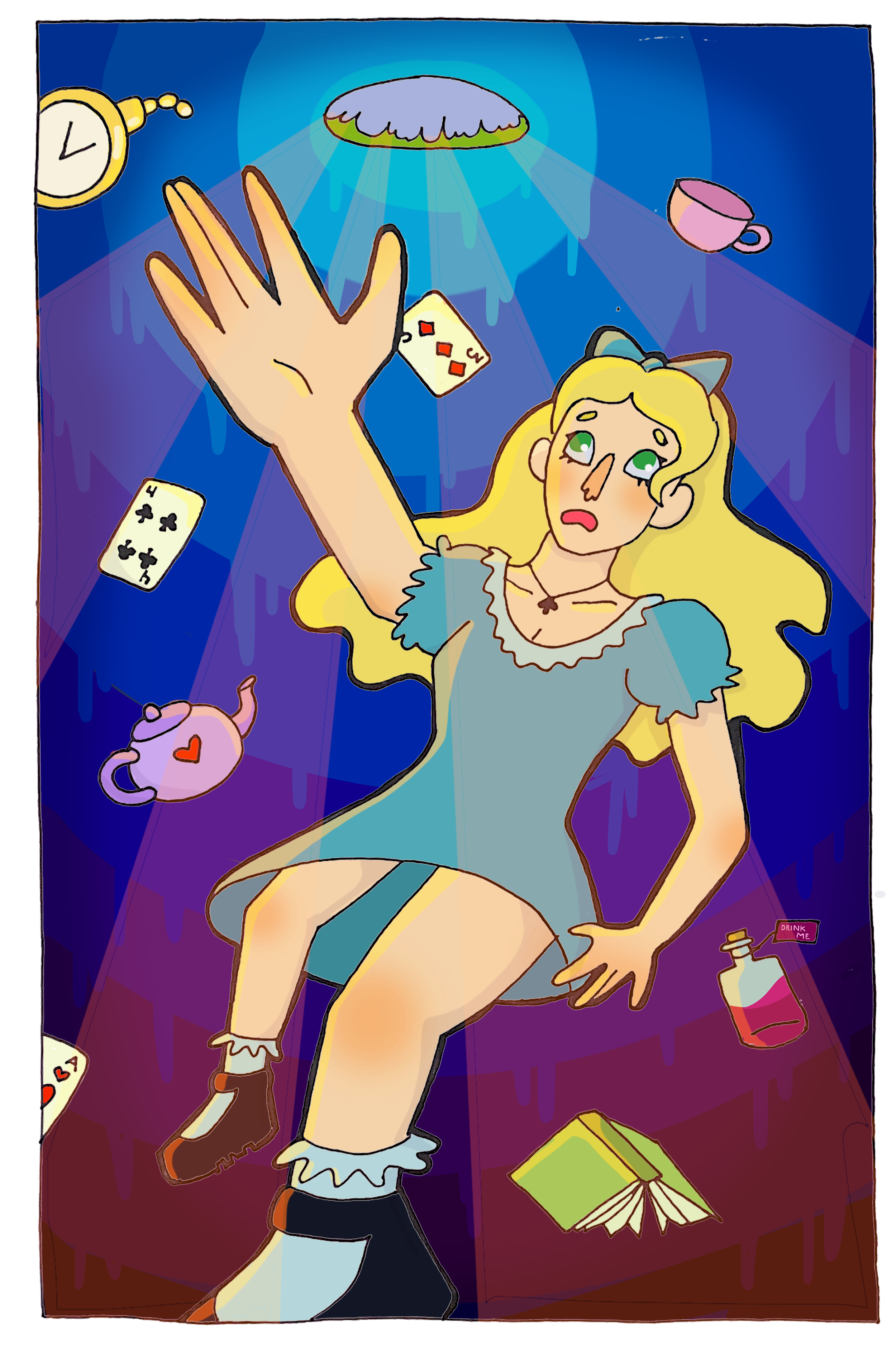
A young girl, age seven, wakes up one more morning to find her limbs have grown dramatically overnight. Her arm, once a mere ten inches, now extends fifteen feet from her body, while her hands have shrunk to a size similar to that of a blueberry. Her leg, once a comfortable few feet from her body, has narrowed to just centimeters in width. Amid her confusion, she recalls a situation similar to her own in a story she once read about a girl who follows a rabbit down a hole: Lewis Caroll’s popular children’s novel, Alice’s Adventures in Wonderland [1]. British psychiatrist Dr. John Todd noticed this same resemblance during the 1950s when six of his adolescent patients came to him complaining of migraines and epileptic episodes, simultaneously reporting symptoms parallel to Alice’s experiences [2].
COMMON SYMPTOMS
Alice in Wonderland Syndrome (AIWS) is a rare neurological condition primarily manifesting in dysmetropsias, anomalies of visual perception in which the shape of an object changes. This can occur in various forms, such as objects seeming further away than they truly are (teleopsia), or closer than they truly are (pelopsia) [2]. Macropsia and micropsia, the perception of objects as larger or smaller than they appear respectively, are the primary diagnostic criteria of AIWS. A greater proportion of individuals believed to have AIWS reported instances involving macropsia and/or micropsia as opposed to other forms of dysmetropsia, though reported symptoms are highly variable [2].
In addition to illusory changes in size and depth, Todd described an intricate group of symptoms associated with AIWS: these included feelings of derealization and depersonalization (feeling separate from oneself), distortions of one’s sense of proprioception (the awareness of the movement and position of one’s body in space), and changes in the patient’s experience of time [2]. As with the principal symptoms of AIWS, they are perceived by the patient rather than observed. Time distortion falls under this category. For AIWS patients, their experience of time can either feel sped-up, or slowed down [3].
FIRST REPORTS
It was John Todd whose analysis of this condition became the most influential in 1955, when he compared the similarities in AIWS symptoms to the experiences of Lewis Caroll’s protagonist, Alice, in the 1865 book, Alice’s Adventures in Wonderland [1][2]. His connection between the aforementioned dysmetropsias and seizures/migraines in relation to Alice’s Adventures in Wonderland gained attention, but this was not the first time this condition may have been described. In 1904, William Spratling noted similar occurrences in his patients who were said to have “seen everything bigger” just before an epileptic seizure [3]. In 1907, William Gowers reported patients seeing things looking “twice the size” during their epileptic episodes, and Hermann Oppenheim’s patients reported “detachments of the trunk or limbs'' during migraines in 1913 [3].
SYMPTOMS, EXPLAINED
Perhaps the most disorienting feature of AIWS is the illusory changes occurring in the patient’s visual field. Despite what the patient themselves may see, there exists no true physical change in the composition of their body. For instance, the unusual phenomenon of their limbs growing very large or very small is a disruption in their visual perception, and should not be confused with a standard hallucination. While both phenomena present a deviation from visual authenticity, hallucinations are experienced regardless of external stimuli, whereas visual disruptions are a product of invalid interpretations by the brain from its incoming sensory and somatosensory inputs [2]. In addition to the lack of hallucinations, AIWS differs from psychosis since patients are aware that what they are seeing is a false interpretation. The commonality between this vast group of symptoms is the constitution of sensory perception distortions, different from standard hallucinations since percepts are experienced without an appropriate external stimulus [2]. The experience of time distortions and feelings of derealization and depersonalization are less commonly studied, as they relate to a more subjective and conscious experience, and this subjective matter makes it difficult to appropriately measure [4].

PATHOLOGY AND PATHOPHYSIOLOGY
Despite the dearth of knowledge pertaining to AIWS’s distinct pathophysiology, edemas of the cerebral regions near the visual pathways of the brain and infectious diseases (occurring before, during, and after onset) may be involved in how AIWS occurs [5]. Since Todd’s first report, AIWS has remained mostly obscure, and in turn, underresearched. Not much information has been available, though increasingly functional approaches in neuroimaging have renewed scientific interest in AIWS. [2]. Few of these studies have suggested specific cortical regions at play in AIWS, but available data points to a region known as the temporoparietaloccipital carrefour (TPO-C) at the junction of the temporal, paritel, and occipital cortices of the brain [6].
In his report, Todd speculated that the temporal lobe, which plays a role in processing visual, emotional, and linguistic perceptions, was involved in AIWS. Further research has suggested the TPO-C is a critical mediator for AIWS, as it is a region in which all somatosensory and visual information is “integrated to create an inner and outer representation of one’s body” [5]. The somatosensory system is the complex network in the body responsible for sensing touch, pain, temperature, body position, and pressure. Sensory neurons relay these feelings from the skin to the brain, and work to aid humans to recognize objects, differentiate between textures, and more.
It generally thought that our senses amount to a collective group of five一audition, taste, touch, smell, and sight. However, the biological system of sense in humans serves as a method of gathering information from the environment around them and appropriately responding to stimuli, and involves a more intricate process of gathering this information. Disordered proprioception, for instance, is one of the key features of AIWS [2]. Though not one of the “main” five senses, proprioception is essential for functioning in everyday life, and to know one’s position in a space is an autonomic process. The TPO-C seems to be integral, not just in potentially mediating AIWS, but for the unification of sensory information into meaningful interpretations. For a patient with AIWS, anomalies possibly occurring here could account for false visual and auditory perceptions, time distortions, and more.
AIWS AND AD SPECTRUM DISORDERS: PROPRIOCEPTION
An increasing interest has formed concerning AIWS’s disturbances in proprioception and coordination. Though the condition remains more of a mystery than a complete answer, patterns of both visual anomalies and proprioceptive anomalies have been of discussion, as well as its potential relationship to AD spectrum disorders [9]. Individuals on the autism spectrum typically have what is known as ‘stimming behaviors’. Stimming is a shortened version of the term “self-stimulatory behavior” and is a repetitive behavior or behaviors such as arm or hand movements and vocal ticking [7]. Stimming is not limited to autism, but is a trademark symptom of and predominantly associated with autism spectrum disorders (ASD) [8]. These behaviors have been widely researchedㅡnumerous studies focused on the autism spectrum have been conducted to understand the mechanisms behind its genetic markers, and to learn why those on the autism spectrum exhibit these behaviors.
It has been found that stimming behaviors not only serve as a self-soothing method, but could possibly occur to compensate for deficits in spatial orientation. In a study performed on autistic children in Hong Kong, the spatial management and orientation of the participants was tested with adjustments to their visual perception and hand-eye coordination [9]. It was found that positive adjustments in their visual perception led to performing better on tasks such as ball-catching, and in some, autonomic motor impulses like their posture [9]. At its core, AIWS is a condition affecting visual perception and proprioception, which integrate to reflect coordination and spatial managament. In some cases of AIWS, spatial deficit-compensation behaviors have been speculated. The commonalities that may lie in these disorienting features has become a topic of discussion, hinting toward a more clear future in understanding the spatial deficits and methods of compensation towards them. Autism and AIWS remain separate conditions, but there exists postulation that the pathology in ASD can provide insight to the neuropathogenesis of AIWS.

DIAGNOSTIC CRITERIA + TREATMENT
AIWS case presentations are highly variable. Thus, symptoms are difficult to measure; an operational definition and objective diagnostic criteria remain unclear and require further expansion. Since its introduction, AIWS has collected 42 visual symptoms and 16 somesthetic symptoms [2], and under 200 cases have been reported [2]. As a result of this diversity of symptoms, alongside the syndrome’s rarity, it is commonly misunderstood and misdiagnosed.
It appears that AIWS is categorized into different classes, distinguished by factors like age, symptoms, and proposed etiologies. Though it has been recorded in patients of varying ages, most of the data comes from juvenile cases [2]. Another factor that complicates accurate diagnosis of this condition is the frequency of spontaneous resolution. That is, it is difficult to draw conclusions about effective clinical treatment because of the condition’s transient nature. While a smaller portion of cases have experienced long-term AIWS without a significant gap between episodes, most cases, which have occurred in young children, tend to resolve on their own as they age without prior recognition or treatment. This ‘random’ onset and rectification may suggest AIWS could occur due to a neurodevelopmental delay, and resolve with further neurodevelopment.
ETIOLOGIES
From its initial reports, AIWS was reported in patients suffering from migraines and/or epilepsy [2]. A variety of other factors, ranging from psychiatric disorders to psychoactive drug induced symptoms, have been associated with the condition, but in adolescent cases especially, infectious diseases are the prevalent etiology for AIWS [5]. Viruses in particular have been of focus, specifically the Epstein-Barr Virus (EBV). EBV remains the predominant viral infection associated with AIWS and features of EBV infections in the central nervous system that may be linked to AIWS consist of diminishing cerebral blood flow, edemas, and encephalitis [5]. Encephalitis is inflammation of the brain, most commonly caused by viral infections, and edemas consist of excess fluid in both the extracellular and intracellular areas of the brain一typically causing increased brain pressure and impairing nerve functions [10][11]. Other viruses, such as the Varicella zoster virus and the H1N1 Influenza A virus [5], have been linked to both the onset and development of AIWS-EBV, however, predominates.
CONCLUSION
Alice in Wonderland Syndrome presents itself very broadly and remains an enigma. The development of neuroimaging technologies has provided a promising future in understanding this intriguing condition and finding effective methods of treatment. Beyond relieving symptoms, the research into sensorimotor integrations and proprioception is building a solid framework for other conditions involving the dysfunction of these processes. The complexity of the brain entangles a multitude of questions and, somewhere, answers yet to be found. With the collective pursuit of these answers, the future of neuroscience remains bright.
References:
[1] Carroll, L., & Tenniel, J. (2015). Alice's adventures in wonderland. London: Macmillan Children's Books.
[2] Blom, J. D. (2016). Alice in wonderland syndrome. Neurology: Clinical Practice, 6(3), 259-270. doi:10.1212/CPJ.0000000000000251
[3] Fine, E., Farooq, O., & Mahfooz, N. (2017). Alice in wonderland syndrome: A history (P2.042). Neurology, 88(16 Supplement), P2.042. Retrieved from http://n.neurology.org/content/88/16_Supplement/P2.042.abstract
[4] Blom, J. D., Nanuashvili, N., & Waters, F. (2021). Time distortions: A systematic review of cases characteristic of alice in wonderland syndrome. Frontiers in Psychiatry, 12, 668633. doi:10.3389/fpsyt.2021.668633
[5] Perez-Garcia, L., Pacheco, O., Delgado-Noguera, L., Motezuma, J. P. M., Sordillo, E. M., & Paniz Mondolfi, A. E. (2021). Infectious causes of alice in wonderland syndrome. Journal of Neurovirology, 27(4), 550-556. doi:10.1007/s13365-021-00988-8 [doi]
[6] Mastria, G., Mancini, V., Viganò, A., & Piero, V. D. (2016). Alice in wonderland syndrome: A clinical and pathophysiological review. BioMed Research International, 2016, undefined-undefined. doi:10.1155/2016/8243145
[7] Shaughnessy, N. (2016). Curious incidents: Pretend play, presence, and performance pedagogies in encounters with autism. In P. Smagorinsky (Ed.), Creativity and community among autism-spectrum youth: Creating positive social updrafts through play and performance (pp. 187-216). New York: Palgrave Macmillan US. doi:10.1057/978-1-137-54797-2_8
[8] Ghanizadeh, A. (2010). Clinical approach to motor stereotypies in autistic children. Iranian Journal of Pediatrics, 20(2), 149-159. Retrieved from PubMed database. Retrieved from https://pubmed.ncbi.nlm.nih.gov/23056697 https://www.ncbi.nlm.nih.gov/pmc/articles/PMC3446025/
[9] Carmody, D., Kaplan, M., & Gaydos, A. (2001). Spatial orientation adjustments in children with autism in hong kong. Child Psychiatry and Human Development, 31, 233-47. doi:10.1023/A:1026481422227
[10] Encephalitis. (n.d.). Www.hopkinsmedicine.org. https://www.hopkinsmedicine.org/health/conditions-and-diseases/encephalitis
[11] Unterberg, A. W., Stover, J., Kress, B., & Kiening, K. L. (2004). Edema and brain trauma. Neuroscience, 129(4), 1019–1027. https://doi.org/10.1016/j.neuroscience.2004.06.046