Neuroprosthetics and the Bionic Human
Author: Stephen Baak || Scientific Reviewer: Gianna Vitelli || Lay Reviewer: Taylor Forry || General Editor: Vishwanka Kuchibhatla || Artists: Matthew Jozwik & Rebecca Gitlevich || Graduate Scientific Reviewer: Michael West
Publication Date: December 20, 2021

Historically, certain conditions have been thought of as untreatable and incurable, such as blindness and paralysis. As a result, any cases of recovery from these conditions were viewed as miraculous and unexplainable. It has not been until recently that mechanisms for many of these conditions have been extensively studied so that treatments have become not only possible, but appear increasingly inevitable.
Through detailed research and experimentation, scientists and engineers have been able to develop treatments for these injuries previously thought to be untreatable. For example, the development of artificial limb interfaces has enabled motion [1] and facilitated sensation [2] in patients with spinal cord injury, while the development of spherical artificial eyes has the potential ability to give sight to the blind [3]. These treatments, which combine natural neuronal processing with prosthetic devices, are referred to as neuroprostheses. Although research for these devices is still ongoing, early results are promising, and may soon give rise to permanent solutions for these conditions.
Manifesting Motion
The spine is not only the primary bone structure that helps us stand upright and support our body, but it serves as the structure that houses the neuronal cells that comprise the spinal cord. The spinal cord is the primary conduit through which information and signals are sent from the brain to the rest of the body, and vice versa [4]. These neuronal cells serve as the wiring that connects the brain to the peripheral nervous system, allowing signals to flow freely throughout the body. The spinal cord extends down from the brain through the cervical spine (neck), below which is the thoracic spine (mid-back), followed by the lumbar spine (lower back), and ultimately the sacral spine (end of the spine).
When there is damage to the spine or spinal cord, the immediate physiological response is swelling and inflammation, causing the spinal cord to expand and occupy the diameter of the spinal canal at the location of the injury [5]. The secondary physiological result is ischaemia, which is the release of toxic chemicals from disrupted neural membranes [5]. This results in a secondary injury cascade that can harm or even kill neighboring cells, exacerbating the initial mechanical damage caused by the injury [5].
Once the spinal cord has been disrupted in this way, nearly all nerve pathways that extend past the point of injury on the spinal cord are inactivated. Signals can be sent from the periphery to the spinal cord, but they cannot bridge the gap that has formed due to the injury. Likewise, signals from the brain cannot overcome this barrier to send information out to the body, resulting in a loss of motor function. This loss of function is what characterizes paralysis, whether it be paraplegia (caused by damage in either the thoracic, lumbar, or sacral regions of the spinal cord) or quadriplegia (caused by damage in the cervical region of the spinal cord) [4].
By studying patterns of spinal cord signaling in pathways that control motor function in the limbs, researchers have been able to develop an artificial signaling system that essentially “bridges the gap” caused by spinal cord injury. Targeted electrical stimulation to individual muscles or groups of muscles can elicit muscle twitches and movement. Activating these muscles artificially to produce individual joint movements is referred to as neuromuscular electrical stimulation [1]. By using different electrical patterns and temporal sequences, researchers have shown that coordinated limb movements are possible in patients with no voluntary motor control in what is referred to as functional electrical stimulation (FES) [1].
Typically, FES has been used as a prosthetic device to assist patients with neurological impairments, such as those recovering from stroke, in performing tasks on their own [1]. Recent developments in brain recording and processing have led to new discoveries and methodologies in brain-computer interface (BCI) technologies. One such development in BCI applications is the ability for these interfaces to translate brain signals from a patient into electrical outputs, which are then used to generate movements using FES [1]. This means that BCI-FES assumes the role of the spinal cord in motor output signalling, allowing the patient to move their limbs on their own again. In addition to the viability of BCI-FES as a permanent prosthetic solution, the use of these technologies in a therapeutic, non-permanent setting has been proven to aid in the recovery of voluntary function [1].
Facilitating Feeling
In the case of BCI-FES in paralyzed patients, the neuroprosthetic devices are implemented in patients that have existing nerve pathways that have been disconnected due to spinal cord injury. In the case of amputees, these pathways have been removed, necessitating the creation of artificial nerve pathways to supplement the remaining portions of these peripheral nerve pathways. These artificial pathways typically serve to facilitate control of the artificial limbs, but recent developments have shown the ability to construct artificial limb interfaces that provide tactile feedback to the user.
Current models have estimated that by the year 2050, approximately 3.6 million Americans will be living with the loss of at least one limb [6]. The primary cause of limb loss in the United States are amputations secondary to dysvascular disease (any abnormal condition of the blood vessels), which accounts for 54% of amputations [6]. The second leading cause of limb loss is loss secondary to trauma, which accounts for about 45% of amputations [6]. In many of these cases, patients elect to utilize prosthetic limbs, although the utility and functionality of many of these limbs is extremely limited when compared to the natural limbs they are replacing. Significant research has been conducted to improve the functionality and control of these artificial limbs, although these brain-machine interfaces primarily serve to send signals from the brain to the artificial limb without any sensory feedback [7].
Previous approaches to produce sensory feedback in artificial limbs have encountered challenges in the specificity of the feedback, as well as barriers to implementation, such as patients experiencing irritation or shock-type sensations [2]. In order to improve the utility of artificial limbs, researchers at the Massachusetts Institute of Technology have developed a cutaneous mechanoneural interface (CMI) to facilitate sensory feedback from artificial limbs [2]. In the CMI model, a muscle is surgically coupled to either a native or regenerative skin flap to enable graded sensations of contact or pressure [2]. In this model, a sensorized prosthetic limb makes contact with a surface and transmits electrical signals to a receiver, which translates these signals into graded electrical impulses that are applied to the skin-muscle structure [2]. Any mechanical deformation of these skin flaps caused by this electrical stimulation is perceived and transduced into sensory signals by free nerve endings and the four low-threshold mechanoreceptors that are present in the skin [2]. These mechanoreceptors generate signals in response to the deformation caused by the electrical stimulation, and the signals generated then travel along the nerve pathways in the remaining portion of the amputated limb, where they ultimately reach the spinal cord and brain, enabling the sensation of touch. Seeing as these signals are being generated by naturally occurring mechanoreceptors in the skin, the sensation is expected to closely mimic natural physiological sensation, although thus far this model has only been constructed in rats [2].
This model is, in theory, applicable for almost all patients who have undergone amputation, no matter the primary cause of the amputation [2]. In patients who have had amputations as a result of injury, joint instability, failed joint stability, or cancer, there is healthy skin tissue available at the site of the amputation that can be used to surgically produce these CMIs [2]. In the case of amputations resulting from severe injury, vascular disease without comorbid diabetes, and congenital limb deficiencies, there is often little to no available healthy skin tissue at the site of amputation [2]. In these cases, the skin tissue necessary for the implementation of the CMI model can be obtained as a skin graft from another part of the patient's body in what is referred to as a “regenerative approach” [2].
While the developers of the CMI model did not design the mechanoneural interface with the express goal of being a potential solution to the phenomenon of phantom limb pain, it may be an unintended benefit of the application of CMI modulated sensitization of prosthetic limbs. Phantom limb pain is a common symptom following amputation, with estimates stating that approximately 80% of all amputation patients experience this phenomenon at some point following their procedure [8]. Phantom limb pain is the feeling of excessive pain originating from limbs that have been amputated, giving the illusion that the limb is still attached to the body [8]. Of the several treatment and therapy options available, some of the non-pharmacological methods include prosthesis training and transcutaneous nerve stimulation, both of which relate to the implementation of the CMI model [8]. While the therapeutic electrical stimulation used as a treatment for phantom limb pain is applied on external skin rather than subcutaneously transplanted skin, the combination of stimulation and prosthetic training with the CMI model may aid in lessening the severity of phantom limb pain in amputation patients.
Synthesizing Sight
Most of us take the use of our eyes for granted, but for a significant portion of the population, their vision is impaired to the point of being blind, whether legally or completely blind. According to estimates, blindness or low vision affects approximately 1 in 28 Americans over the age of 40, and this number is expected to increase with the aging population [9]. While there have been previous devices designed to enable the visually impaired and blind to see, recent developments in nanoscale technology have enabled the development of spherical artificial eyes with the potential ability to give full sight to the blind without the need for an external apparatus.
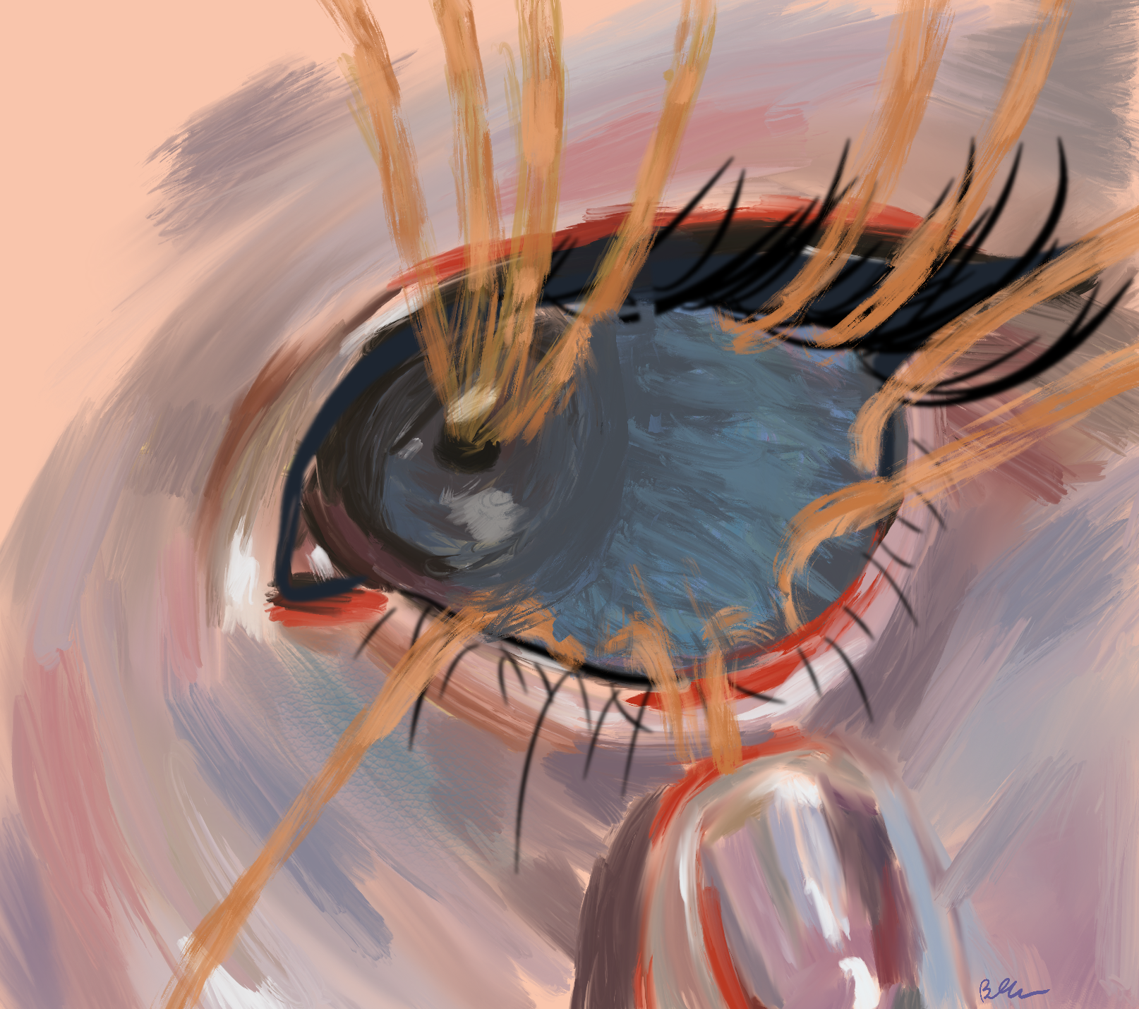
The human eye is composed of several layers and structures, although the parts primarily responsible for vision are the cornea, pupil, lens, retina, and optic nerve [10]. Light enters the eye through the cornea, which is the front layer of the eye, and passes through the pupil, which is the black opening in the center [10]. The pupil is expanded and contracted by the iris in response to the intensity of light, allowing either more or less light to enter the eye [10]. After light passes through the pupil, it goes through the lens and vitreous humor, which adjusts to bend and focus the light onto the retina, which is located in the back of the eye [10]. The retina contains specialized structures called photoreceptors, which are made up of rods and cones [10]. Rods allow us to see shapes, while cones function to allow us to see colors [10]. These rods and cones translate this information into electrical signals, which are transmitted through the optic nerve to the occipital lobe located in the back of the brain [10].
Blindness can be caused by a dysfunction or failure in any part of the aforementioned visual pathway. Some of the most pressing causes of blindness are retinal degenerative diseases such as retinitis pigmentosa and age-related macular degeneration [11]. Previous research has worked toward restoring a rudimentary form of vision in patients who are blind, but these methods are unable to restore full vision [11]. These previous methods follow the same general outline in terms of design, with both external and internal modules [11]. The external module typically consists of a camera affixed to the patient, usually designed as a pair of glasses or goggles, an external computer to process the image, and a wireless interface to transmit the processed information to the internal module [11]. The internal module is implanted into the patient and consists of an embedded controller and a back-end stimulator. The controller receives signals from the external module and transforms this information into electrical stimulation patterns that the back-end stimulator administers to the target tissue of the retina [10]. These stimulation patterns mimic the natural signalling that takes place in the eye and thus are able to be interpreted by the nervous system and result in the formation of rudimentary images that are visible to the patient [11].
Current visual prosthesis produce images that are often lacking in resolution and are without a wide field of view (FOV), which stems from the use of cameras and similar flat image sensors to serve as light detectors rather than biomimetic ocular devices [12]. Devices that mimic the human eye’s ability to detect and perceive light are highly desirable not only for image capture, but also as a potential form of artificial eyes for the blind [12]. Previous attempts to develop hemispherical retinas for biomimetic eyes have been limited due to the planar fabrication processes of their components [12]. To improve upon this method, researchers at the Hong Kong University of Science and Technology have successfully developed a spherical biomimetic electrochemical eye, known as the EC-EYE [12]. The EC-EYE has been constructed using a hemispherical template, with light-sensing nanowires constructed directly onto the hemispherical surface rather than being constructed on a planar surface and manipulated into a hemispherical shape [12]. These light-sensing nanowires function as the artificial form of rods and cones found in the biological eye [12]. The spherical shape of the EC-EYE is completed with a lens and artificial iris at the front of the device joined to the hemispherical retina at the back of the device, and the entire spherical arrangement is filled with an ionic fluid that mimics the vitreous humor [3].
What is striking about the EC-EYE is that its hemispherical retina not only allows for nanowires that are more densely packed than those found in other devices, but for a higher photoreceptor density than that found in the human eye [3]. In the human eye, the density of photoreceptors is about 10⁷ photoreceptors per square centimeter, while in the EC-EYE the nanowire density is about 4 x 10⁸ wires per square centimeter, almost 50 times more dense than the biological eye [3]. As a result of this increased density, it is possible that the EC-EYE is capable of achieving higher image resolution than the biological eye, while the use of liquid-metal wires behind the sensing material mean that the EC-EYE avoids the blind-spot issue that occurs in the human retina [11].
The EC-EYE still has several flaws that need to be addressed, namely the costly manufacturing method, the need for nanowires to be reduced in size to increase resolution and reduce scale, and the necessity for more rigorous durability testing [3]. Additionally, the method of administration of the EC-EYE as a prosthetic device poses several questions, as the EC-EYE would replace the existing eyes of a patient. There are several side-effects with removing eyes, namely potential disruption of the circadian rhythm, which is the internal clock of the body [13]. Despite these drawbacks, the successful development of a spherical artificial eye is an important proof-of-concept. This means that the development of spherical artificial eyes for prosthetic implementation is a development that is certainly possible in the near future [3].
Science Fiction Into Science Fact
For years, whether in writing, movies, or television, people have been fascinated by the possibility of merging man and machine. From the series The Six Million Dollar Man to the Star Wars saga, the idea of technology combining with mankind has been a concept reserved for science-fiction. Now, with the improvement of modern technology and research methods, the devices seen in these stories are no longer fictional, and with continuous developments, may soon become a part of everyday life.
References:
Milosevic, M., Marquez-Chin, C., Masani, K., Hirata, M., Nomura, T., Popovic, M. R., & Nakazawa, K. (2020). Why brain-controlled neuroprosthetics matter: Mechanisms underlying electrical stimulation of muscles and nerves in rehabilitation. BioMedical Engineering OnLine, 19(1). https://doi.org/10.1186/s12938-020-00824-w
S. Srinivasan, S., & M. Herr, H. (2021). A cutaneous mechanoneural interface for neuroprosthetic feedback. Nature Biomedical Engineering. https://doi.org/10.1038/s41551-020-00669-7
Jiang, H. (2020). Artificial eye boosted by hemispherical retina. Nature, 581(7808), 264–265. https://doi.org/10.1038/d41586-020-01420-7
Kirshblum, S. C., Burns, S. P., Biering-Sorensen, F., Donovan, W., Graves, D. E., Jha, A., Johansen, M., Jones, L., Krassioukov, A., Mulcahey, M., Schmidt-Read, M., & Waring, W. (2011). International standards for neurological classification of spinal cord injury (Revised 2011). The Journal of Spinal Cord Medicine, 34(6), 535–546. https://doi.org/10.1179/204577211x13207446293695
McDonald, J. W., & Sadowsky, C. (2002). Spinal-cord injury. The Lancet, 359(9304), 417–425. https://doi.org/10.1016/s0140-6736(02)07603-1
Ziegler-Graham, K., MacKenzie, E. J., Ephraim, P. L., Travison, T. G., & Brookmeyer, R. (2008). Estimating the prevalence of limb loss in the United States: 2005 to 2050. Archives of Physical Medicine and Rehabilitation, 89(3), 422–429. https://doi.org/10.1016/j.apmr.2007.11.005
Adewole, D. O., Serruya, M. D., Harris, J. P., Burrell, J. C., Petrov, D., Chen, H. I., Wolf, J. A., & Cullen, D. K. (2016). The evolution of neuroprosthetic interfaces. Critical Reviews in Biomedical Engineering, 44(1–02), 123–152. https://doi.org/10.1615/critrevbiomedeng.2016017198
Flor, H. (2002). Phantom-limb pain: Characteristics, causes, and treatment. The Lancet Neurology, 1(3), 182–189. https://doi.org/10.1016/s1474-4422(02)00074-1
Causes and prevalence of visual impairment among adults in the United States. (2004). Archives of Ophthalmology, 122(4), 477. https://doi.org/10.1001/archopht.122.4.477
Forrester, J. V., Xu, H., Kuffová, L., Dick, A. D., & McMenamin, P. G. (2010). Dendritic cell physiology and function in the eye. Immunological Reviews, 234(1), 282–304. https://doi.org/10.1111/j.0105-2896.2009.00873.x
Maghami, M. H., Sodagar, A. M., Lashay, A., & Riazi-Esfahani, H. (2014). Visual prostheses: The enabling technology to give sight to the blind. Journal of Ophthalmic and Vision Research, 9(4), 494. https://doi.org/10.4103/2008-322X.150830
Gu, L., Poddar, S., Lin, Y., Long, Z., Zhang, D., Zhang, Q., Shu, L., Qiu, X., Kam, M., Javey, A., & Fan, Z. (2020). A biomimetic eye with a hemispherical perovskite nanowire array retina. Nature, 581(7808), 278–282. https://doi.org/10.1038/s41586-020-2285-x
Lickey, M. E., Wozniak, J. A., Block, G. D., Hudson, D. J., & Augter, G. K. (1977). The consequences of eye removal for the circadian rhythm of behavioral activity in Aplysia. Journal of Comparative Physiology ? A, 118(1), 121–143. https://doi.org/10.1007/bf00612342