A Labyrinth of Connection: The Link Between Social Exclusion And Academic Performance in Adolescents
Author: Heta Darji || Scientific Reviewer: Gabija Stewart || Lay Reviewer: Anika Bhatnagar || General Editor: Julia Barth
Artist: Sumita Nagendra || Graduate Scientific Reviewer: Dana Zeid
Publication Date: December 19th, 2025
Introduction
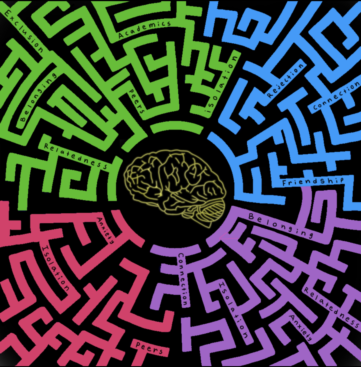
We often think of school as a place to socialize, grow, and develop unique parts of our identity. But for some, what if it were a place of social isolation? Social exclusion in school, an environment swarmed by peers, can leave a devastating impact on children. If seen from a broader perspective, events like social exclusion and rejection can negatively impact a child’s brain biology [1]. As the impacted brain regions play a role in said child’s education, a change in academic performance can occur as well [1, 2].
While belonging and connectedness are important at any age, these needs are particularly relevant in adolescence. Research suggests that adolescents are highly susceptible to peer rejection. As a group, they may experience significant mental health effects in response, including depression and anxiety. Adolescence is typically a time of increased independence from family and heavy dependence on peers, which may hold some explanation as to why it is so crucial for development [3,4].
Parts of the Brain Involved in Social Interaction
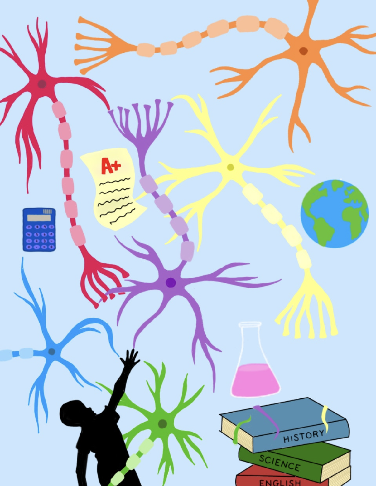
The brain is the “control center” of the nervous system, controlling various processes including emotions, movement, memory, and appetite. Alongside those functions, different parts of the brain work together to interpret social interactions [5].
Although it is relatively small, the hypothalamus is recognized as a control center that maintains the body in a state of homeostasis, or balance. It is located near the brainstem or the base of the brain. To perform its function, the hypothalamus produces hormones. Hormones are signaling molecules that ultimately allow for the relaying of a biological “message” (such as the awareness of hunger) to distant organs. Hormones are generally steroids (lipid-derived) or proteins (made of amino acids). In the context of the Hypothalamic-Pituitary-Adrenal (HPA) axis, the hypothalamus first releases hormones into the system of capillaries surrounding the pituitary gland, which is located below the hypothalamus [6]. These signals direct the anterior pituitary (one of the two lobes of the pituitary gland) to release hormones of its own to reach a desired effect. An important hormone the hypothalamus releases is the corticotropin-releasing hormone (CRH), which stimulates the pituitary to secrete adrenocorticotropic hormone (ACTH). ACTH goes on to stimulate the adrenal glands located on top of the kidneys [7]. These glands play a crucial role in the stress response. In response to ACTH, the adrenal glands secrete the stress hormone cortisol [8].
This pathway between the hypothalamus, pituitary gland, and adrenal gland is known as the Hypothalamic-Pituitary-Adrenal (HPA) axis, which regulates cortisol levels to enable humans to respond quickly to threats. Studies on cortisol have found that levels usually spike soon after waking up and start to decline throughout the day. This wasn’t the case in socially excluded children, though. Their morning cortisol levels were higher, and there was less of the expected decline of cortisol over the course of the day compared to socially included children [1]. Healthy HPA axis functioning is linked to better academic performance in children because of the support it provides to adjusting in school [9].
This elevation of cortisol also impacts another part of the brain important in social interaction: the hippocampus. This brain region plays a vital role in the formation, storage, and retrieval of memory [10]. Research has shown that the hippocampus enables people to utilize their imagination and recall past social interactions through memory. This can result in the person displaying empathy and viewing events from a perspective different from their own [11]. Additionally, the hippocampus contributes to communication. During social interactions, words are pulled from memory, processed, and given meaning to relay information. Access to past knowledge and the creation of new sentences enable humans to get points across during conversation [12, 13, 14]. These complex functions showcase the hippocampus’s abilities beyond memory formation [13].
Increased general cortisol levels also affect the hippocampus [15]. A study done on 44 children showcased how memory functions under stress. Researchers used a popular memory game and stressors to find that stressed children demonstrated higher levels of cortisol. They also performed poorly in the memory games, suggesting that cortisol can impact memory retrieval [16]. Learning and memory are crucial for the successful education of children, as those with better memory generally perform better academically. In contrast, those who struggle with memorization often see it manifest in their academic performance [17].
Another part of the brain crucial for social interaction is the amygdala, or amygdalae, which are located next to the hippocampi. This region is known for its involvement in the fear response, but its function extends to aggression, learning, addictive behaviors, and social interactions. Furthermore, the amygdala aids in interpreting the intent of a peer through their language and behavior. Multiple psychological and neurological conditions and disorders also impact the amygdala, such as anxiety, post-traumatic stress disorder, phobias, etc. [18, 19].
To carry out its function, the amygdala also reacts to stressors like social exclusion. Data show that individuals with high levels of stress have increased amygdala activity. Similarly, socially excluded individuals have increased amygdala reactivity [20]. This increase can contribute to reduced emotion regulation and negative emotional experiences [21]. For students, emotional management has a strong correlation to academic success. It allows students to stay focused and motivated throughout the various social and academic struggles in school, keeping them invested in their learning [22].
Conclusion
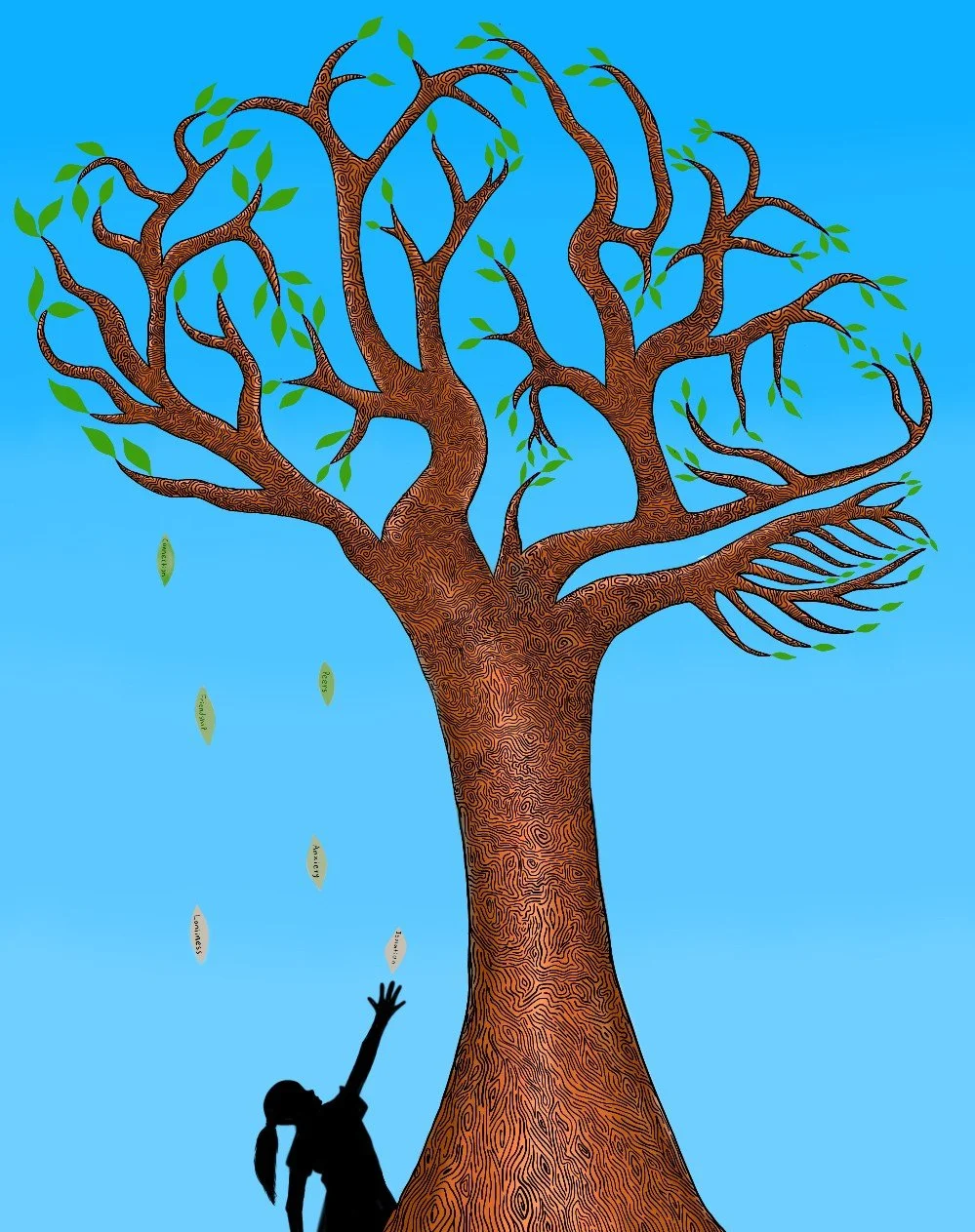
Social exclusion is more than just an unfortunate event; it’s a brain-altering experience. In summary, social exclusion can cause cortisol increases that dysregulate HPA axis functioning, disrupt memory formation in the hippocampus, and heighten the sensitivity of the amygdala. The impact of childhood social exclusion on brain regions involved in stress, memory, and social processing can hinder the academic development of students.
Such links between ideas in literature can be the basis for better preventative measures, hopefully limiting social exclusion’s detrimental effects on the adolescent brain. This can be seen as more teacher interference when peer rejection occurs, and the development of skills like empathy, stress management, and confidence that are needed for students to work through social challenges healthily.
References
Peters, E. J., Riksen-Walraven, M., Cillessen, A. H. N., & De Weerth, C. (2011). Peer rejection and HPA activity in middle childhood: Friendship makes a difference. Child Development, 82(6), 1906–1920. https://doi.org/10.1111/j.1467-8624.2011.01647.x
Burdakov, D., & Peleg-Raibstein, D. (2020). The hypothalamus as a primary indicator of memory updating. Physiology & Behavior, 223, 112988. https://doi.org/10.1016/j.physbeh.2020.112988
Cross, D., Shaw, T., Hearn, L., Epstein, M., Monks, H., Lester, L., & Thomas, L. (2009). Australian covert bullying prevalence study (ACBPS). Child Health Promotion Research Centre, Edith Cowan University. https://ro.ecu.edu.au/ecuworks/6795/
Leets, L., & Sunwolf. (2005). Adolescent rules for social exclusion: When is it fair to exclude someone else? Journal of Moral Education, 34(3), 343–362. https://doi.org/10.1080/03057240500211618
Adkins-Regan, E. (2009). Neuroendocrinology of social behavior. ILAR Journal, 50(1), 5–14. https://doi.org/10.1093/ilar.50.1.5
Saper, C. B., & Lowell, B. B. (2014). The hypothalamus. Current Biology, 24(23), R1111–R1116. https://doi.org/10.1016/j.cub.2014.10.023
Megha, R., Wehrle, C. J., Kashyap, S., & Leslie, S. W. (2022). Anatomy, abdomen and pelvis: Adrenal glands (suprarenal glands). In StatPearls. StatPearls Publishing. https://www.ncbi.nlm.nih.gov/books/NBK482264/
Herman, J. P., McKlveen, J. M., Ghosal, S., Kopp, B., Wulsin, A., Makinson, R., Scheimann, J., & Myers, B. (2016). Regulation of the hypothalamic–pituitary–adrenocortical stress response. Comprehensive Physiology, 6(2), 603–621. https://doi.org/10.1002/cphy.c150015
Graham, A. M., Pears, K. C., Kim, H. K., Bruce, J., & Fisher, P. A. (2018). Effects of a school readiness intervention on hypothalamus–pituitary–adrenal axis functioning and school adjustment for children in foster care. Development and Psychopathology, 30(2), 419–431. https://doi.org/10.1017/S0954579417001171
Anand, K. S., & Dhikav, V. (2012). Hippocampus in health and disease: An overview. Annals of Indian Academy of Neurology, 15(4), 239–246. https://doi.org/10.4103/0972-2327.104323
Beadle, J. N., Sheehan, A. H., Dahlben, B., & Gutchess, A. H. (2015). Aging, empathy, and prosociality. The Journals of Gerontology: Series B, Psychological Sciences and Social Sciences, 70(2), 215–224. https://doi.org/10.1093/geronb/gbt091
Lemke, J. L. (2000). Across the scales of time: Artifacts, activities, and meanings in ecosocial systems. Mind, Culture, and Activity, 7(4), 273–290. https://doi.org/10.1207/S15327884MCA0704_03
Rubin, R. D., Watson, P. D., Duff, M. C., & Cohen, N. J. (2014). The role of the hippocampus in flexible cognition and social behavior. Frontiers in Human Neuroscience, 8, 742. https://doi.org/10.3389/fnhum.2014.00742
Cacioppo, J. T., Hughes, M. E., Waite, L. J., Hawkley, L. C., & Thisted, R. A. (2006). Loneliness as a specific risk factor for depressive symptoms: Cross-sectional and longitudinal analyses. Psychology of Aging, 21(1), 140–151. https://doi.org/10.1037/0882-7974.21.1.140
Lammer, L., Beyer, F., Luppa, M., Sanders, C., Baber, R., Engel, C., Wirkner, K., Löffler, M., Riedel-Heller, S. G., Villringer, A., & Witte, V. (2023). Impact of social isolation on grey matter structure and cognitive functions: A population-based longitudinal neuroimaging study. eLife, 12, e83660. https://doi.org/10.7554/eLife.83660
Quesada, A. A., Wiemers, U. S., Schoofs, D., & Wolf, O. T. (2012). Psychosocial stress exposure impairs memory retrieval in children. Psychoneuroendocrinology, 37(1), 125–136. https://doi.org/10.1016/j.psyneuen.2011.05.013
Gathercole, S. E., Lamont, E., & Alloway, T. P. (2006). Working memory in the classroom. In Working memory and education (pp. 219–240). Academic Press. https://doi.org/10.1016/B978-012554465-8/50010-7
Stevens, J. S., & Jovanovic, T. (2018). Role of social cognition in post-traumatic stress disorder: A review and meta-analysis. Genes, Brain and Behavior, 18(1), e12518. https://doi.org/10.1111/gbb.12518
Gongora, M., Teixeira, S., Martins, L., Marinho, V., Velasques, B., Moraes, L., Nicoliche, E., Bastos, V. H., Nunes, M. K., Cartier, C., Nascimento, V., Vicente, R., Silva, L. W. D. G., de Carvalho, M., Di Giacomo, J., Junqueira, J., Santos, F., Cagy, M., Oliveira, T., & Ribeiro, P. (2019). Neurobiological evidences, functional and emotional aspects associated with the amygdala: From “what is it?” to “what’s to be done?”. Journal of Neuropsychiatry, 9(3), 2379–2396. https://www.jneuropsychiatry.org/peer-review/neurobiological-evidences-functional-and-emotional-aspects-associated-with-the-amygdala-from-what-is-it-to-whats-to-be-d.pdf
Clark, U. S., Miller, E. R., & Hegde, R. R. (2018). Experiences of discrimination are associated with greater resting amygdala activity and functional connectivity. Biological Psychiatry: Cognitive Neuroscience and Neuroimaging, 3(4), 367–378. https://doi.org/10.1016/j.bpsc.2017.11.011
Gerin, M. I., Viding, E., Pingault, J.-B., Puetz, V. B., Knodt, A. R., Radtke, S. R., Brigidi, B. D., Swartz, J. R., Hariri, A. R., & McCrory, E. J. (2019). Heightened amygdala reactivity and increased stress generation predict internalizing symptoms in adults following childhood maltreatment. Journal of Child Psychology and Psychiatry, 60(7), 752–761. https://doi.org/10.1111/jcpp.13041
Valiente, C., Swanson, J., & Eisenberg, N. (2012). Linking students’ emotions and academic achievement: When and why emotions matter. Child Development Perspectives, 6(2), 129–135. https://doi.org/10.1111/j.1750-8606.2011.00192.x