The Hidden Price of Glory: Investigating How Sports-Related Head Trauma Increases the Risk of Brain Disease
Author: Saad Farooqui || Scientific Reviewer: Sulaiman Sharief || Lay Reviewer: Pranesh Rajamuthiah || General Editor: Alicia Hoffmann
Artist: Raquel Roberge || Graduate Scientific Reviewer: Claire Deckers
Publication Date: December 19th, 2025
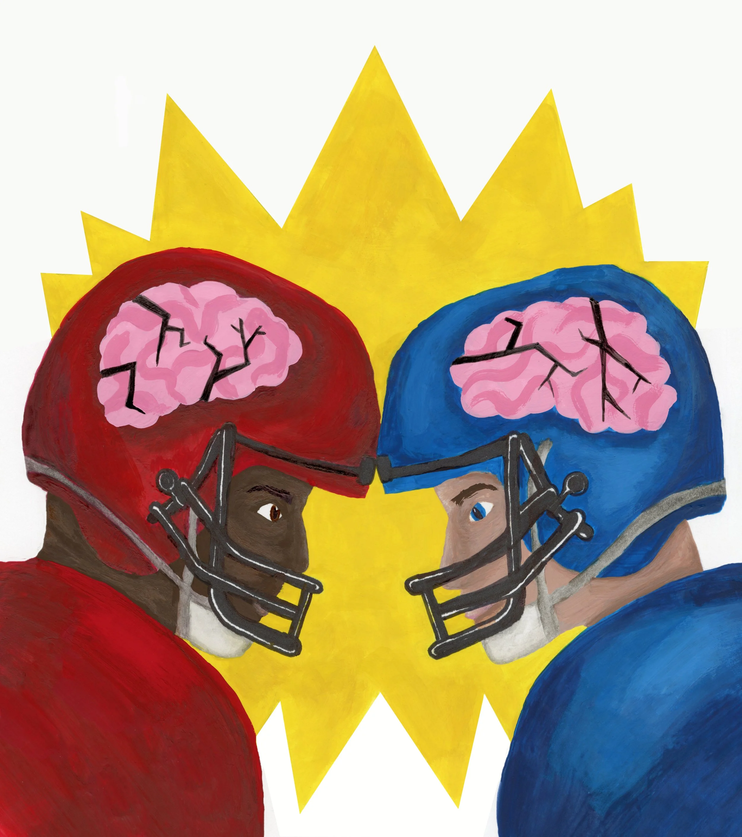
It’s the dark side of the game: the hard hits, the glory, and the hidden cost carried long after the cheering stops. For years, athletes and fans of all sports have shrugged off concussions as the price of admission, but a growing body of research suggests that the violence inherent in contact sports is writing a tragic final chapter for some of its biggest stars.
A traumatic brain injury (TBI) occurs when an individual suffers an injury to the head, often through blunt force or a fall. This is often caused by the brain rapidly shifting forward and backward inside the skull, creating what are known as coup/contrecoup injuries. This trauma can cause a wide range of effects, from mild headaches to life-threatening injuries [1]. Individuals with TBI often suffer from brain damage, which can lead to neurological deficits. These initial physical impacts are considered primary TBI effects, but they often trigger devastating secondary effects that worsen over time. Acute effects of TBI might subside, but research suggests that injury can still increase susceptibility to frontotemporal dementia (FTD) [2]. This condition is notorious for drastically changing a person's personality, behavior, and language skills—essentially altering who they are. TBIs have been shown to cause traumatic axonal injury (TAI), which increases amyloid plaque buildup [3]. This buildup involves sticky clumps of abnormal protein that collect outside the brain's nerve cells, effectively jamming the lines of communication between neurons. This severe disruption is a crucial hallmark of escalating neurodegenerative damage. Abnormal protein activity is a common pathological finding in many neurodegenerative disorders, including FTD andAlzheimer's Disease (AD) [4]. Specifically, TBI is believed to trigger two distinct pathological mechanisms that accelerate neurodegenerative disease: one leading to the amyloid-plaque accumulation of AD, and another associated with the tau-based protein pathology of FTD. This is particularly concerning for individuals who participate in contact sports, such as American football or hockey.
These clinical findings are tragically visible in the real world. Many former professional athletes, particularly football players, have had their final years defined not by touchdowns or glory, but by confusing behavioral shifts, memory loss, and severe emotional instability. The long-term neurological fallout rewrites their life script, showing the consequences of repeated head injury (RHI) and the urgent need for a solution.
Researchers recruited 264 participants, half of whom were FTD patients and half healthy controls, carefully matching them by age and sex to investigate whether head trauma accelerates the disease. The team collected detailed histories on the frequency and severity of TBI to pinpoint how head trauma influenced the age when FTD symptoms first appeared. The data is clear: the frequency of head trauma correlates with premature FTD onset.
Many of these behavioral symptoms observed are also hallmarks of FTD; to investigate this link, a recent study analyzed participants from separate FTD memory care facilities [2]. The researchers first assessed participants based on their method of TBI, measuring the duration of loss of consciousness (LOC), the method of injury, the frequency of TBI, and the age at which the TBI occurred [2]. The researchers found that the frequency of TBI with LOC had a negative linear correlation with the onset of FTD symptoms. This indicated a clear relationship—more TBI exposure means a younger FTD onset. The average onset of FTD symptoms in patients without TBI was 63 years old, compared to an average of 55 years in patients with two or more TBI. The researchers also found that the healthy controls played an average of two years of American football, in contrast to the FTD sample, who played an average of five years of American football [2]. In short, they found that the longer someone played football, the earlier they developed FTD symptoms. These results suggest that repeated TBI in addition to head impacts from contact sports where RHI risk is elevated are correlated and may be a cause of FTD.
To understand the specific mechanism by which TBI increases AD risk, the amyloid pathway must be examined. The initial blunt-force trauma causes TAI. TAI immediately boosts the production of beta-amyloid protein [2]. These sticky amyloid plaques then accumulate outside the neurons, which disrupts communication between nerve cells and drives neurotoxicity. This process initiates the progressive degeneration characteristic of AD [5]. Research suggests a strong link between a single, moderate-to-severe TBI and the later onset of AD, indicating that the mechanical injury fundamentally changes the brain's protein environment.
To understand the actual mechanism by which TBIs may lead to neurodegeneration, it is necessary to consider the abnormal protein deposits involved. TBIs are thought to increase the risk of neurodegenerative disease in part due to the traumatic axonal injury (TAI) that occurs after the damage [3]. The TAI produces an increase in beta-amyloid protein buildup, known as amyloid plaques, which in turn can lead to further neurotoxicity and degeneration [6]. It is important to note that while FTD and Alzheimer's Disease (AD) are distinct disorders, they do share some similar behavioral and pathological features, allowing for relevant comparative studies. Another study sought to analyze the connection between TBIs and amyloid plaques [3]. Twenty-eight participants, 9 with TBI, 10 with AD, and 9 controls, were assessed based on beta-amyloid buildup. The study's results showed a positive correlation between TBI and TAI in TBI patients compared to the AD and control groups. This suggests that amyloid plaque buildup is correlated with TAI resulting from TBI.
Pathological changes, such as the clumping of abnormal proteins like tau and TDP-43, are key features of FTD. In FTD, these proteins often clump in the frontal and temporal regions of the brain. This clumping causes loss of function, which accounts for the onset of FTD. The loss of function often includes neuronal death, which leads to problems with coordination, language, and executive function [6]. The next study employed imaging and statistical analysis to investigate amyloid ligands and the correlation between FTD and beta-amyloid proteins in post-mortem brains [7]. The results of this experiment showed a strong presence of beta-amyloid proteins in the white matter (deeper stem-like structures of the neurons in the brain) of FTD brains (positive correlation), in contrast to an insignificant correlation of beta-amyloid in the pons, cerebellum, and medulla. The positive correlation suggests that the increase in beta amyloid protein in the white matter of the cortex may cause FTD.
The Bigger Picture
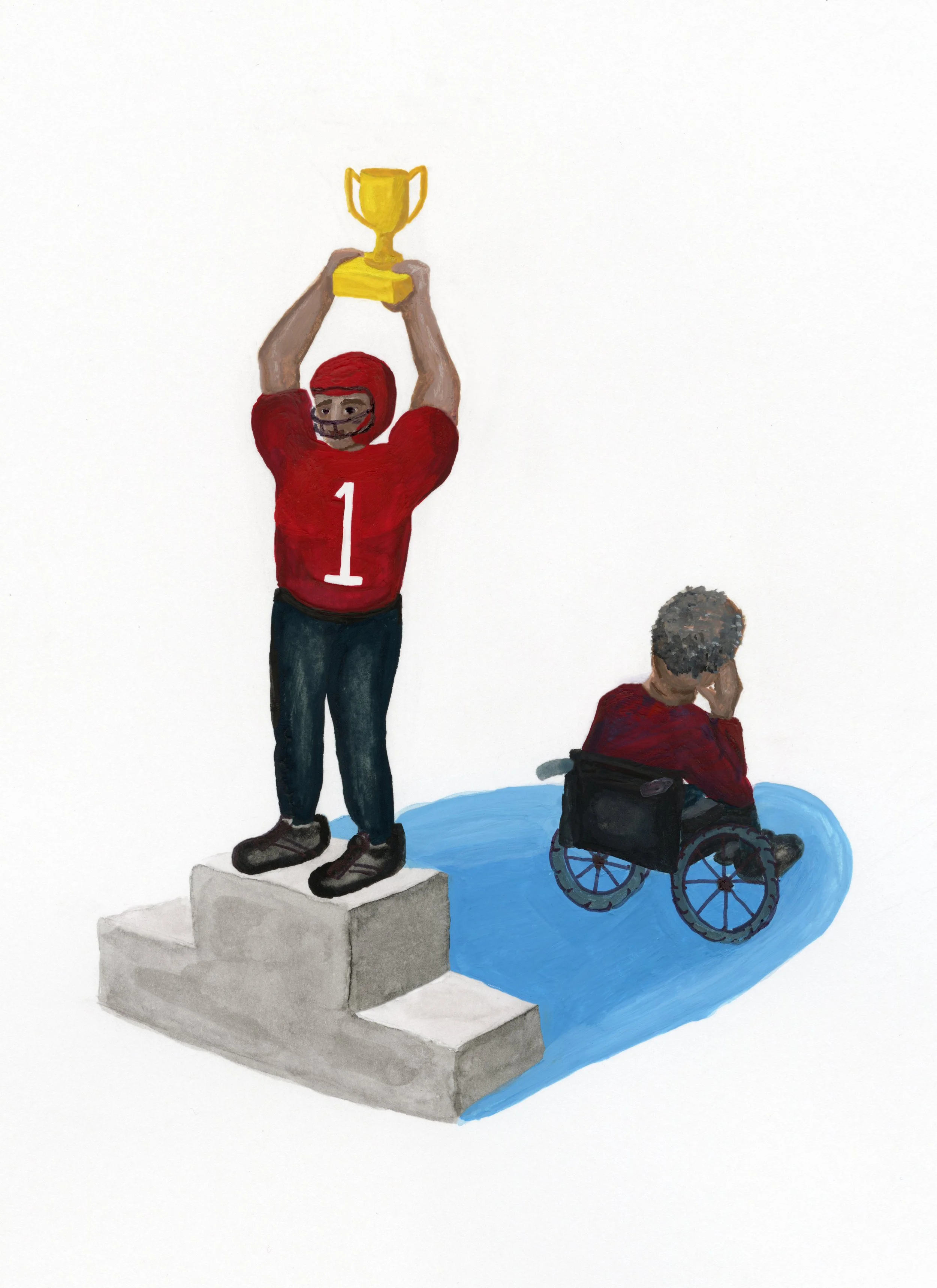
The most alarming takeaway from these findings is the clear acceleration of FTD onset caused by repetitive sports trauma. The data suggests a potent dose-response relationship: the more frequently an individual experiences head impacts, and the longer they participate in high-contact sports, the younger they are when the disease begins to manifest. Moving the average onset age by several years—from the early sixties down to the mid-fifties—presents a stark warning that the chronic physical stress of the game is actively shortening the functional lifespan of the brain, making the neurological cost undeniable.
The combined findings paint a clear picture: repeated TBI is strongly linked to the creation of abnormal protein buildup, and higher amounts of this abnormal protein seem to accelerate FTD symptoms. This protein trouble, which is far more prevalent in TBI patients than in healthy individuals, seems to accelerate FTD symptoms. Specifically, more TBIs are shown to lead to memory problems, and the buildup of these plaques is directly associated with FTD itself. Taken together this suggests the brain changes triggered by a blow to the head are a key factor in the development of the disease.
At the center of this problem is the traumatic axonal injury (TAI) caused by head impact, which appears to be the mechanism that drives FTD. The evidence from studies analyzing high-impact sports constantly shows a spike in this beta-amyloid protein damage, which helps explain why the pursuit of athletic glory comes with such a significant price. Understanding this cost to athletes' long-term health is vital. Finding the definite cause of FTD could dramatically improve millions of lives every year, particularly those who have participated in contact sports. However, based on the limitations we've discussed, more precise studies are urgently needed to determine if there is a clear causative relationship (TBI definitely causes FTD) rather than just a strong link (correlative relationship).
References
National Institute of Neurological Disorders and Stroke. (n.d.). Traumatic brain injury (TBI).https://www.ninds.nih.gov/health-information/disorders/traumatic-brain-injury
Asken, B. M., Bove, J. M., Bauer, R. M., Tanner, J. A., Casaletto, K. B., Staffaroni, A. M., VandeVrede, L., Alosco, M. L., Mez, J. B., Stern, R. A., Miller, B. L., Grinberg, L. T., Boxer, A. L., Gorno-Tempini, M. L., Rosen, H. J., Rabinovici, G. D., & Kramer, J. H. (2024). Clinical implications of head trauma in frontotemporal dementia and primary progressive aphasia. Alzheimer’s Research & Therapy, 16, 193. https://doi.org/10.1186/s13195-024-01432-1
Holmes, B. B., et al. (2025). β-Amyloid induces microglial expression of GPC4 and APOE leading to increased neuronal tau pathology and toxicity. bioRxiv, 2025.02.20.637701. https://doi.org/10.1101/2025.02.20.637701
Zhang, Y., Schuff, N., Du, A.-T., Rosen, H. J., Kramer, J. H., Gorno-Tempini, M. L., Miller, B. L., & Weiner, M. W. (2009). White matter damage in frontotemporal dementia and Alzheimer’s disease measured by diffusion MRI. Brain, 132(9), 2579–2592. https://doi.org/10.1093/brain/awp071
Kent, S. A., Spires-Jones, T. L., & Durrant, C. S. (2020). The physiological roles of tau and Aβ: Implications for Alzheimer’s disease pathology and therapeutics. Acta Neuropathologica, 140(4), 417–447.https://doi.org/10.1007/s00401-020-02196-w
Scott, G., Ramlackhansingh, A. F., Edison, P., Hellyer, P., Cole, J., Veronese, M., Leech, R., Greenwood, R. J., Turkheimer, F. E., Gentleman, S. M., Heckemann, R. A., Matthews, P. M., Brooks, D. J., & Sharp, D. J. (2016). Amyloid pathology and axonal injury after brain trauma. Neurology, 86(9), 821–828. https://doi.org/10.1212/WNL.0000000000002413
Tan, R. H., Kril, J. J., Yang, Y., Tom, N., Hodges, J. R., Villemagne, V. L., Rowe, C. C., Leyton, C. E., Kwok, J. B. J., Ittner, L. M., & Halliday, G. M. (2017). Assessment of amyloid β in pathologically confirmed frontotemporal dementia syndromes. Alzheimer’s & Dementia: Diagnosis, Assessment & Disease Monitoring, 9, 10–20. https://doi.org/10.1016/j.dadm.2017.06.004