Parkinson’s Petri Dish: What Fruit Flies Teach Us About Brain Disease
Author: Sarah Bhanushali || Scientific Reviewer: Sai Karanam || Lay Reviewer: Lillian Hubbard || General Editor: Shardul Garg
Artist: Devin Palmer || Graduate Scientific Reviewer: Marina Kaplan
Publication Date: December 19th, 2025
Introduction: The Fly that Changed the Brain
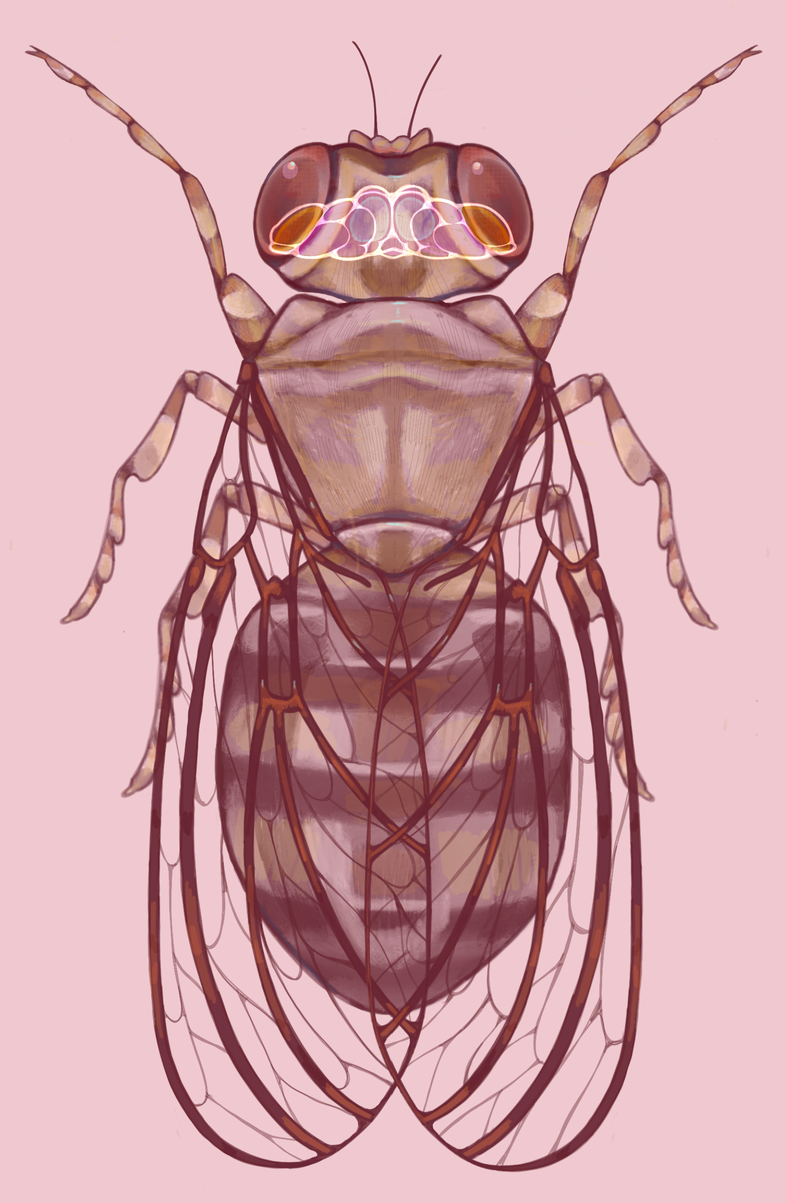
Parkinson’s disease (PD) research is not always conducted in humans or even mice. Sometimes, it is buzzing around in a plastic tube with red eyes and wings.
They live around 50 to 80 days, and they are the size of a sesame seed [1]. Yet, they have helped decode one of the most devastating diseases of the brain in our time: Parkinson’s. Drosophila melanogaster, the fruit fly, has reshaped and transformed neuroscience research. Although it may seem insignificant, this tiny insect has become a powerhouse model organism, revealing the genetic roots of Parkinson’s disease. Drosophila models have played a crucial role in advancing Parkinson’s disease research, contributing to key discoveries in Parkinson’s genes, mitochondrial dysfunction, and new treatments [2].
What is Parkinson's Disease (PD)?
Parkinson’s disease (PD) affects over 10 million people globally, a number projected to reach 25.2 million by 2050 [3, 4]. It is linked to abnormal buildup and misfolding of a protein called α-synuclein in the brain [5]. These protein clumps, known as Lewy bodies, contribute to neuronal dysfunction and cell death, leading to progressive neurodegeneration. Once neurodegeneration has spread to the substantia nigra (SN), a structure at the base of the brainstem, patients typically experience motor symptoms such as bradykinesia (slowness of movement), muscular rigidity, tremor, and postural instability [6]. The SN produces dopamine, a chemical messenger important for communication between the brain and the body [7]. When neurodegeneration occurs in the SN, this leads to less production of dopamine and ultimately results in loss of coordinated, smooth purposeful movement.
Although no definitive cure for PD currently exists, there are several methods of treatment including levodopa, dopamine agonists, MAO-B inhibitors, along with physical therapy to improve balance and mobility. In more advanced cases, surgical procedures like deep brain stimulation (DBS), may be performed [8]. While most cases of PD arise without a known cause, a significant subset is linked to mutations in specific genes. This has driven researchers to use model organisms, like Drosophila, to uncover the molecular pathways behind the disease [9].
Why Use Drosophila in Brain Research
Studying the human brain directly is challenging, and animal models can be time-consuming and costly. Roughly 75% of human disease-related genes have Drosophila counterparts, making Drosophila valuable for modeling human biology [10]. Despite having only around 140,000 neurons compared to 86 billion in humans, they share dopaminergic circuits affected in PD [11]. When Drosophila lose dopaminergic neurons, they struggle to climb or fly, showing clear and quantifiable signs of motor dysfunction that mirror human symptoms of PD [2].
Drosophila are also compatible with advanced genetic techniques, including CRISPR/Cas9 and RNAi, which allow precise manipulation of gene expression and modeling of disease-related mutations [12]. Using these tools, researchers can switch specific genes on or off or introduce Parkinson’s-related mutations that mirror those found in human patients, then observe how these changes disrupt cellular pathways in the brain. This genetic flexibility makes Drosophila an efficient platform for high-throughput drug screening, allowing a large number of potential therapies to be tested rapidly and simultaneously [13].
Additionally, Drosophila have a rapid reproductive rate, which enables multi-generation studies in just weeks. This makes it possible to track how genetic mutations or treatments affect offspring over time. Combined with their low maintenance cost and minimal space requirements, Drosophila models are highly efficient for large-scale, time-sensitive research [14].
Key Genetic Discoveries from Drosophila Models
PINK1 and Parkin:
A major breakthrough in PD research involved PINK1 (PTEN-induced kinase 1) and Parkin, two specific genes linked to early-onset PD [15]. Researchers discovered that there were specific mutations on these genes often found in PD patients but their functions in the brain were not entirely understood. The PINK1 protein acts as a sensor that detects any damaged mitochondria, while the Parkin protein works to repair damaged mitochondria by tagging them for removal, a process called mitophagy. The PINK1-Parkin pathway is crucial for maintaining mitochondrial health. Together, they act as a quality-control system for the mitochondria, which is where energy is produced. Drosophila are useful in studying how damage to this pathway contributes to the onset of PD [16].
In a 2006 study, researchers discovered removing PINK1 weakened muscles and damaged mitochondria, causing difficulties in movement and abnormal wing posture. However, when researchers increased expression of Parkin in these PINK1-deficient flies, a majority of their problems improved, suggesting that Parkin overexpression can rescue cells impacted by neurodegeneration and reverse muscle dysfunction. Conversely, when researchers increased PINK1 expression in flies that lacked Parkin, their problems did not improve [17]. This study demonstrated the important role of the PINK1-Parkin pathway in regulating mitochondrial function which established mitochondrial dysfunction as a central mechanism of PD.
α-synuclein:
The protein α-synuclein is predominantly found in the brain and is involved in neurotransmitter release and cell communication. When α-synuclein misfolds and clumps together, Lewy bodies form. They interfere with crucial cellular processes, leading to synaptic dysfunction, mitochondrial damage, disrupted protein degradation, and even death of dopaminergic neurons [18, 19].
Researchers have genetically engineered Drosophila in order to study the development of Lewy bodies and the loss of dopaminergic neurons, which resulted in decreased locomotor ability. These effects demonstrate the neurodegeneration that occurs in PD patients and how this manifests clinically. Studying this process in Drosophila provided a fast, genetically tractable way to confirm the central role of α-synuclein in Parkinson’s pathology [20].
LRRK2 and DJ-1:
Mutations in LRRK2 (Leucine-rich repeat kinase 2) are the most common genetic cause of PD. The LRRK2 gene produces a protein that adds phosphate groups to other proteins, a process called phosphorylation. This helps regulate several cellular processes and is crucial to neuronal homeostasis [21]. In a 2008 study, researchers mutated the LRRK2 gene in Drosophila to model Parkinson’s and to assess the effects of levodopa, the most effective drug for PD to date. Results showed that locomotor functions improved in Drosophila, but the treatment did not prevent the loss of neurons important for motor function. This study helped clarify the role of LRRK2 mutations in PD and informed later development of LRRK2-targeted therapies to treat PD [22].
The PARK-7 gene is commonly associated with autosomal recessive, early-onset parkinsonism (AREP) [23]. A mutation on this gene can result in the altered structure and function of the DJ-1 protein which is primarily known for its role in neuroprotection and response to oxidative stress, the body’s ability to neutralize the unstable molecules (reactive oxygen species) produced by the mitochondria during normal cellular respiration. Oxidative stress contributes to further mitochondrial dysfunction, formation of Lewy bodies, and death of dopaminergic neurons [24]. Mitochondrial dysfunction can occur due to various genetic and environmental factors, such as neurotoxins [25]. In a 2005 study, researchers knocked out the DJ-1 gene in Drosophila. This resulted in hypersensitivity to environmental neurotoxins, leading to rapid degeneration of the flies’ dopaminergic neurons [26]. This study of Drosophila demonstrated the complex interplay of environment and genetics in PD.
What Flies Teach Us About the Parkinson’s Brain
Mitochondrial quality control:
A clear example came from studying PINK1/Parkin. In flies, loss of PINK1 led to damaged mitochondria and movement problems. When researchers increased Parkin, many of these problems improved. This showed that neurons need a working system to find and remove damaged mitochondria, and that Parkin acts after PINK1 to carry out this job [17]. Later studies in human PD cells reported similar problems with mitophagy, which showed that a pathway first mapped in flies is also present in human material [27]. In other words, the fly model helped identify the order of events in a disease pathway that matters in human PD patients.
Different PD genes create the same kind of cellular stress:
Research in Drosophila has shown that very different Parkinson’s-related mutations, such as those affecting α-synuclein, LRRK2, and DJ-1, all converge on the same problem: oxidative stress and overwhelmed cellular cleanup systems. Drosophila that overexpress α-synuclein develop Lewy body-like protein clumps, mitochondrial dysfunction, and loss of dopaminergic neurons, mirroring what is seen in human PD tissue [28]. Similarly, Drosophila with LRRK2 mutations show impaired autophagy and reduced neuron survival, while Drosophila lacking DJ-1 are extremely sensitive to oxidative stress and rapidly lose dopaminergic neurons [29]. These studies demonstrated different pathways from which PD-causing mutations ultimately damage mitochondria, overload protein-clearing systems, and push neurons into the same toxic stress state. This insight helped researchers recognize oxidative stress as a central, unifying mechanism of PD and informed later studies on mitochondrial protection and protein-clearance therapies in human models.
An accelerated view of a slow disease:
While PD is characterized by insidious onset in humans, the Drosophila’s rapid generation cycle and short lifespan provide many advantages in PD research. In Drosophila, removing one protective step, such as mitophagy, autophagy, or oxidative defense, can be enough to cause neuron loss within their short lifespan. In humans, the same processes fail, but more gradually. This manifests as inherited mutations, age-related decline of cellular cleanup, and increasing protein aggregation [30]. Therefore, Drosophila provide an accelerated model of PD that makes the sequence easier to see: toxic cellular byproducts build up, resulting in cellular dysfunction in dopaminergic neurons.
The Power of Tiny: Using Fruit Flies to Find Treatment
High-throughput Drug Screening:
Researchers use Drosophila to rapidly test hundreds of therapeutic compounds for their ability to protect dopaminergic neurons. For instance, the use of vitamin E or the SOD1 gene can help reverse degeneration in PINK1-deficient flies. Their antioxidant properties help neutralize harmful reactive oxygen species and prevent cellular damage that would otherwise lead to neuron loss [31]. In another screen, the compound zaprinast improved movement in DJ-1 mutant flies and protected human neuron-like cells as it had the most significant ability to reduce oxidative stress [32]. These studies show how Drosophila models can allow researchers to efficiently identify the most impactful disease-modifying drugs for PD.
Gene Therapy:
Researchers test genetic therapies in Drosophila before transitioning to more sophisticated mammal models. Using tools like RNA interference (RNAi) to silence harmful genes, or CRISPR to correct mutations, they can restore cellular pathways in fly models of Parkinson’s. Repairing these pathways helps limit the mitochondrial damage and protein buildup that drives degeneration [33]. These experiments provide early proof-of-concept that gene-targeted therapies could modify disease progression rather than just manage symptoms. Other gene-based tests include knocking down overactive LRRK2 with RNAi to reduce its toxicity, boosting autophagy genes to help clear α-synuclein aggregates, and overexpressing antioxidant genes such as SOD1 to better handle oxidative stress [33, 34]. All of these showed at least partial protection in Drosophila, which supported gene-targeted approaches that can modify the disease process, not just the symptoms.
Behavioral Rescue:
One of the major strengths of Drosophila models is that improvements can be seen directly in behavior. Drosophila with Parkinson’s-related mutations often show climbing defects or reduced activity. In humans, PD is marked by slowness of movement and problems with balance and posture; in flies, the slower climbing and reduced locomotion are close equivalents to those motor symptoms [35]. When researchers restore the affected pathway, the behavior improves. Loss of PINK1 in Drosophila caused wing and locomotor defects. Increasing Parkin in those same flies reduced the mitochondrial damage and the flies regained their ability to move normally [17]. This recovery happened because more dopaminergic neurons were preserved, which translated to movement as a whole.

Limitations of Fly Models
Although Drosophila have revealed major insights into Parkinson’s, they are not exact replicas of humans. Their brains are far less complex and lack structures, like the basal ganglia, which are central to human movement. They cannot mimic many nonmotor symptoms such as mood changes or cognitive decline [30]. The course of disease looks very different from what occurs in humans due to a shorter life span and simple nervous system. Therefore, Drosophila studies are just the first step. After researchers identify important genes or potential treatments in Drosophila, they confirm those findings with further research in animals and human stem-cell–derived neurons. Using this chain of models helps connect the basic insights from flies to the full range of symptoms patients experience [27].
Conclusion
Drosophila may be small, but their scientific impact is immense. Drosophila studies have changed how researchers think about PD by revealing how damaged mitochondria, toxic protein clumps, and oxidative stress drive neurodegeneration. Research in Drosophila models has also helped identify potential therapies for cell restoration and improving clinical symptoms.
Although they cannot model every human symptom, their genetic similarities and speed make them indispensable in PD research. Each discovery in Drosophila lays the groundwork for breakthroughs in more complex systems, propelling science closer to therapies that protect neurons and improve quality of life. In the end, Drosophila have been and will continue to be vital to understanding a disease that affects millions of people worldwide.
References
Yusuf, A. O., Danborno, B., Bauchi, Z. M., Sani, D., & Ndams, I. S. (2024). Aging impaired locomotor and biochemical activities in Drosophila melanogaster Oregon R (Fruit fly) model. Experimental Gerontology, 197, 112593. https://doi.org/10.1016/j.exger.2024.112593
Muñoz-Soriano, V., & Paricio, N. (2011). Drosophila Models of Parkinson’s Disease: Discovering Relevant Pathways and Novel Therapeutic Strategies. Parkinson’s Disease, 2011, 1–14. https://doi.org/10.4061/2011/520640
American Parkinson's Disease Association. (2017). What is Parkinson’s? | American Parkinson Disease Association APDA. https://www.apdaparkinson.org/what-is-parkinsons/
Su, D., Cui, Y., He, C., Yin, P., Bai, R., Zhu, J., Lam, J. S. T., Zhang, J., Yan, R., Zheng, X., Wu, J., Zhao, D., Wang, A., Zhou, M., & Feng, T. (2025). Projections for prevalence of Parkinson’s disease and its driving factors in 195 countries and territories to 2050: Modelling study of Global Burden of Disease Study 2021. BMJ, 388, e080952. https://doi.org/10.1136/bmj-2024-080952
Kouli, A., Torsney, K. M., & Kuan, W.-L. (2018). Parkinson’s disease: Etiology, neuropathology, and pathogenesis. In T. B. Stoker & J. C. Greenland (Eds.), Parkinson’s Disease: Pathogenesis and Clinical Aspects. Codon Publications. http://www.ncbi.nlm.nih.gov/books/NBK536722/
Berg, D., Postuma, R. B., Bloem, B., Chan, P., Dubois, B., Gasser, T., Goetz, C. G., Halliday, G. M., Hardy, J., Lang, A. E., Litvan, I., Marek, K., Obeso, J., Oertel, W., Olanow, C. W., Poewe, W., Stern, M., & Deuschl, G. (2014). Time to redefine pd? Introductory statement of the mds task force on the definition of Parkinson’s disease. Movement Disorders, 29(4), 454–462. https://doi.org/10.1002/mds.25844
National Institute of Neurological Disorders and Stroke (NINDS). (2025). Parkinson’s disease. National Institute of Neurological Disorders and Stroke https://www.ninds.nih.gov/health-information/disorders/parkinsons-disease
Zahoor, I., Shafi, A., & Haq, E. (2018). Pharmacological treatment of Parkinson’s disease. In T. B. Stoker & J. C. Greenland (Eds.), Parkinson’s Disease: Pathogenesis and Clinical Aspects. Codon Publications. http://www.ncbi.nlm.nih.gov/books/NBK536726/
Xiong, Y., & Yu, J. (2018). Modeling Parkinson’s disease in Drosophila: What have we learned for dominant traits? Frontiers in Neurology, 9, 228. https://doi.org/10.3389/fneur.2018.00228
University of Cambridge. (2015). How close are you to a fruit fly? University of Cambridge.https://www.cam.ac.uk/research/news/how-close-are-you-to-a-fruit-fly
Doctrow, B. (2024). Complete wiring map of an adult fruit fly brain. National Institutes of Health (NIH). https://www.nih.gov/news-events/nih-research-matters/complete-wiring-map-adult-fruit-fly-brain
Xue, Z., Wu, M., Wen, K., Ren, M., Long, L., Zhang, X., & Gao, G. (2014). CRISPR/Cas9 mediates efficient conditional mutagenesis in Drosophila. G3: Genes|Genomes|Genetics, 4(11), 2167–2173. https://doi.org/10.1534/g3.114.014159
Jennings, B. H. (2011). Drosophila – a versatile model in biology & medicine. Materials Today, 14(5), 190–195. https://doi.org/10.1016/s1369-7021(11)70113-4
Tolwinski, N. S. (2017). Introduction: Drosophila—a model system for developmental biology. Journal of Developmental Biology, 5(3), 9. https://doi.org/10.3390/jdb5030009
Dumitrescu, E., Copeland, J. M., & Venton, B. J. (2023). Parkin knockdown modulates dopamine release in the central complex, but not the mushroom body heel, of aging Drosophila. ACS Chemical Neuroscience, 14(2), 198–208. https://doi.org/10.1021/acschemneuro.2c00277
Von Stockum, S., Nardin, A., Schrepfer, E., & Ziviani, E. (2016). Mitochondrial dynamics and mitophagy in Parkinson’s disease: A fly point of view. Neurobiology of Disease, 90, 58–67. https://doi.org/10.1016/j.nbd.2015.11.002
Clark, I. E., Dodson, M. W., Jiang, C., Cao, J. H., Huh, J. R., Seol, J. H., Yoo, S. J., Hay, B. A., & Guo, M. (2006). Drosophila pink1 is required for mitochondrial function and interacts genetically with parkin. Nature, 441(7097), 1162–1166. https://doi.org/10.1038/nature04779
Stefanis, L. (2012). Α-synuclein in Parkinson’s disease. Cold Spring Harbor Perspectives in Medicine, 2(2), a009399. https://doi.org/10.1101/cshperspect.a009399
Suzuki, M., Sango, K., & Nagai, Y. (2022). Roles of α-synuclein and disease-associated factors in Drosophila models of Parkinson’s disease. International Journal of Molecular Sciences, 23(3), 1519. https://doi.org/10.3390/ijms23031519
Moore, J., Wu, T., Dhindsa, J., El Fadel, O., Le, A., Perez, A., Amoh, B., Tarkunde, A., Zhu, K. F., Avalos, M., Dammer, E. B., Duong, D. M., Seyfried, N. T., Shulman, J. M., Al-Ramahi, I., & Botas, J. (2025). Longitudinal multi-omics in alpha-synuclein Drosophila model discriminates disease- from age-associated pathologies in Parkinson’s disease. Npj Parkinson’s Disease, 11(1), 46. https://doi.org/10.1038/s41531-025-00899-z
Jeong, G. R., & Lee, B. D. (2020). Pathological functions of LRRK2 in Parkinson’s disease. Cells, 9(12), 2565. https://doi.org/10.3390/cells9122565
Liu, Z., Wang, X., Yu, Y., Li, X., Wang, T., Jiang, H., Ren, Q., Jiao, Y., Sawa, A., Moran, T., Ross, C. A., Montell, C., & Smith, W. W. (2008). A Drosophila model for LRRK2-linked parkinsonism. Proceedings of the National Academy of Sciences of the United States of America, 105(7), 2693–2698. https://doi.org/10.1073/pnas.0708452105
Bonifati, V., Rizzu, P., Squitieri, F., Krieger, E., Vanacore, N., van Swieten, J. C., Brice, A., van Duijn, C. M., Oostra, B., Meco, G., & Heutink, P. (2003). DJ-1(Park7), a novel gene for autosomal recessive, early onset parkinsonism. Neurological Sciences, 24(3), 159–160. https://doi.org/10.1007/s10072-003-0108-0
Repici, M., & Giorgini, F. (2019). DJ-1 in Parkinson’s disease: Clinical insights and therapeutic perspectives. Journal of Clinical Medicine, 8(9), 1377. https://doi.org/10.3390/jcm8091377
Blesa, J., Trigo-Damas, I., Quiroga-Varela, A., & Jackson-Lewis, V. R. (2015). Oxidative stress and Parkinson's disease. Frontiers in Neuroanatomy, 9. https://doi.org/10.3389/fnana.2015.00091
Meulener, M., Whitworth, A. J., Armstrong-Gold, C. E., Rizzu, P., Heutink, P., Wes, P. D., Pallanck, L. J., & Bonini, N. M. (2005). Drosophila DJ-1 mutants are selectively sensitive to environmental toxins associated with Parkinson's disease. Current Biology, 15(17), 1572–1577. https://doi.org/10.1016/j.cub.2005.07.064
Hsieh, C.-H., Shaltouki, A., Gonzalez, A. E., Bettencourt da Cruz, A., Burbulla, L. F., St Lawrence, E., Schüle, B., Krainc, D., Palmer, T. D., & Wang, X. (2016). Functional impairment in miro degradation and mitophagy is a shared feature in familial and sporadic Parkinson's disease. Cell Stem Cell, 19(6), 709–724. https://doi.org/10.1016/j.stem.2016.08.002
Zhou, W., Schaack, J., Zawada, W. M., & Freed, C. R. (2002). Overexpression of human α-synuclein causes dopamine neuron death in primary human mesencephalic culture. Brain Research, 926(1), 42–50. https://doi.org/10.1016/S0006-8993(01)03292-9
Yang, Y., Gehrke, S., Haque, Md. E., Imai, Y., Kosek, J., Yang, L., Beal, M. F., Nishimura, I., Wakamatsu, K., Ito, S., Takahashi, R., & Lu, B. (2005). Inactivation of Drosophila DJ-1 leads to impairments of oxidative stress response and phosphatidylinositol 3-kinase/Akt signaling. Proceedings of the National Academy of Sciences, 102(38), 13670–13675. https://doi.org/10.1073/pnas.0504610102
Shadrina, M., & Slominsky, P. (2021). Modeling Parkinson's disease: Not only rodents? Frontiers in Aging Neuroscience, 13. https://doi.org/10.3389/fnagi.2021.695718
Wang, D., Qian, L., Xiong, H., Liu, J., Neckameyer, W. S., Oldham, S., Xia, K., Wang, J., Bodmer, R., & Zhang, Z. (2006). Antioxidants protect PINK1-dependent dopaminergic neurons in Drosophila. Proceedings of the National Academy of Sciences, 103(36), 13520–13525. https://doi.org/10.1073/pnas.0604661103
Sanz, F. J., Solana-Manrique, C., Torres, J., Masiá, E., Vicent, M. J., & Paricio, N. (2021). A high-throughput chemical screen in DJ-1β mutant flies identifies zaprinast as a potential Parkinson's disease treatment. Neurotherapeutics, 18(4), 2565–2578. https://doi.org/10.1007/s13311-021-01134-2
Victor Atoki, A., Aja, P. M., Shinkafi, T. S., Ondari, E. N., Adeniyi, A. I., Fasogbon, I. V., Dangana, R. S., Shehu, U. U., & Akin-Adewumi, A. (n.d.). Exploring the versatility of Drosophila melanogaster as a model organism in biomedical research: A comprehensive review. Fly, 19(1), 2420453. https://doi.org/10.1080/19336934.2024.2420453
Sarkar, S., & Feany, M. B. (2021). Precision medicine on the fly: Using Drosophila to decipher gene-environment interactions in Parkinson's disease. Toxicological Sciences, 182(2), 159–167. https://doi.org/10.1093/toxsci/kfab060
Dabool, L., Juravlev, L., Hakim-Mishnaevski, K., & Kurant, E. (2019). Modeling Parkinson’s disease in adult Drosophila. Journal of Neuroscience Methods, 311, 89–94. https://doi.org/10.1016/j.jneumeth.2018.10.018